Research Article
Isolation and Characterization of Silver Nanoparticles in Cardiospermum halicacabum l. Leaf Extract
Hema J1, Gayathri V1, Sanandam J1 and Anitha R1
1Department of Botany, Avinashilingam Institute for Home Science and Higher Education for Women, Coimbatore, India
2Department of Botany, Bharathi Women’s College (Autonomous), Chennai, India
*Corresponding author: Gayathri V, Department of Botany, Avinashilingam Institute for Home Science and Higher
Education for Women, Coimbatore, India; E-mail: gayathri_bot@avinuty.ac.in
Copyright: © Hema J, et al. 2022. This is an open access article distributed under the Creative Commons Attribution License, which
permits unrestricted use, distribution, and reproduction in any medium, provided the original work is properly cited.
Article Information: Submission: 12/07/2022; Accepted: 10/08/2022; Published: 13/08/2022
Abstract
Cardiospermum halicacabum L. is a medicinal plant with enormous therapeutic properties. The synthesis and characterization of silver nanoparticles
using Cardiospermum halicacabum extract was studied. The synthesis of silver nanoparticles was confirmed by a change in the colour within 10 to 15 min.
Green synthesis of silver nanoparticles is an economically viable approach. The visual observations, EDAX and SEM spectroscopic techniques confirmed
the formation of silver nanoparticles.
Keywords
Cardiospermum; EDAX; Green synthesis; Nano particles; SEM
Introduction
Nanotechnology is a field that is burgeoning day by day and
making an impact in all spheres of human life. Nature and its products
lead to the growth of advancements in the synthesis of nanoparticles.
In the modern field of material science, Nanotechnology is one of
the upcoming fields. This field is an interdisciplinary science that
includes physics, chemistry, biology, material science and medicine.
Nano particles are particles between 1 to 100 nm in size [1]. At
molecular level, using engineered nanodevices and nanostructures,
nanomedicine is being applied in monitoring, repair, construction and
control of human biological systems. Nanotechnology applications
are highly suitable for biological molecules, because of their exclusive
properties [2]. Metal nano particles have a high specific surface area
and a high fraction of surface atoms. A variety of techniques for silver
nanoparticle synthesis have been reported by Iravani et al. [3].
The silver nanoparticles are usually synthesized by reducing silver
salts with the help of reducing agents from biological sources [4].
The silver nanoparticles are synthesized by chemical, physical and
biological methods. The chemical and physical methods for synthesis of nanoparticles are costly and release toxic by-products in nature.
Due to these problems, biological method is an alternative source for
the synthesis of silver nanoparticles [1]. Earlier, the use of biological
methods of silver nanoparticle synthesis using biological entities like
bacteria, fungi and plants has been reported.
The use of biological entities like bacteria, plant extracts or plant
biomass for the production of nanoparticles could be an eco-friendly
approach. Microorganisms trap metal ions from the environment that
are converted into nanoparticles by an enzymatic process. Roy (2017)
reported the synthesis of silver nanoparticles from medicinal plants.
Cardiospermum halicacabum L. (Sapindaceae), commonly called
the balloon vine, is a weed found throughout the country. It finds its
place in traditional systems of healing. It has enormous medicinal
activities, such as diuretic, rubefacient, analgesic, anti-inflammatory
activity, vasodepressant activity, antispasmodic, antirheumatism [5].
Medicinally important weeds can be used effectively in the production
of nanoparticles . Hence, in this study, an attempt has been made to
synthesize silver nanomaterial with Cardiospermum halicacabum.
Material & Methods
In the present study, leaf extract of Cardiospermum halicacabum
L. was taken for green synthesis and characterization of silver
nanoparticles.
Collection of plant samples:
The fresh and healthy leaves of Cardiospermum halicacabum were
collected from an area near Kallakurichi, Tamil Nadu, India.Morphology of the plant:
Cardiospermum halicacabum L.
Systematic position
Kingdom : Plantae
Class : Angiosperms
Order : Sapindales
Family : Sapindaceae
Genus : Cardiospermum
Species : C. halicacabum L.Common names: Karodiyo, Kagdodiyo, Balloon Vine and Heartseed.
Botanical description:
➢ Tropical and sub-tropical, annual and perennial, slender and
beautifully delicate climber- with flower-peduncle tendrils.
➢ Cardiospermum halicacabum is a deciduous climbing shrub
growing about 3 meters tall (Figure 1).
➢ The stems scramble over the ground, climbing into the
surrounding vegetation. Leaves are twice ternate, segments
lanceolate, serrate and acute. Umbellate cyme inflorescence.
➢ Sepals 4, imbricate, outer smaller, inner larger. Petals 4,
rounded at apex. Ovary 3-celled, style very short, trifid.
Medicinal uses:
➢ The entire balloon vine plant possesses several therapeutic
properties.
➢ It is a good diuretic, diaphoretic, emmenagogue and laxative.
➢ Balloon vine is employed for treating stiffness of the limbs,
rheumatism and snakebites.Preparation of Leaf Powder:
The leaves of the medicinal plant taken for the present study were
collected, cleaned and air dried, under shade for about three weeks.
After drying, the leaves are blended using a household electric blender.
This fine powder was analyzed for SNPs characterization study.Green synthesis of silver nanoparticles using leaf extract:
For the synthesis of silver nanoparticles, 1mM aqueous solution
of silver nitrate was prepared and used. 10ml of plant extract was
mixed with 90ml of aqueous 1mM AgNo3 solution in a 250 ml sterile
Erlenmeyer flask. The solution was heated for a few seconds. Later, the
reduction of silver ions was observed by a change in the colour of the
solution from yellow to brown.Characterization of Silver Nanoparticles:
The characterization of silver nanoparticles was carried out by
different techniques.SEM Analysis:
Scanning Electron Microscopy is a commonly used method for
characterization of silver nanoparticles. A thin film of sample was
prepared on a carbon coated grid by placing a very small amount
of sample on the grid, and then it was allowed to dry and examined
under SEM.EDAX analysis:
The sputtering of the sample was done using a SC7620 Sputter
Coater unit under the nitrogen atmosphere. A small strip of carbon
tape was stuck on an aluminium stub and a pinch of sample was
placed on the carbon tape. The elemental analysis and distribution of
the particles were carried out using EDAX with a SUTW-SAPHIRE
model detector.Results & Discussion
The results of the green synthesis and characterization studies on
silver nanoparticles showed the following results.
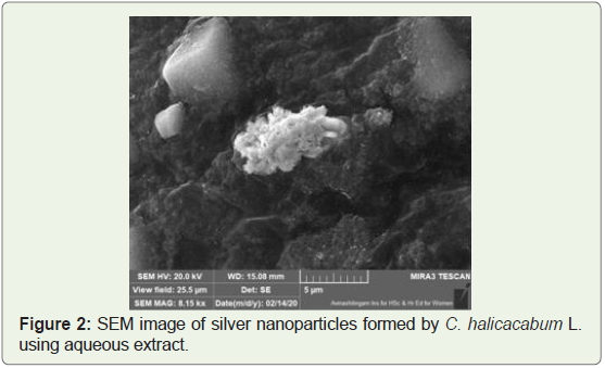
Scanning Electron Microscope:
In Scanning Electron Microscope (SEM) analysis, high resolution
images are generated by focusing a high energy beam of electrons on
the surface of the specimen. These electrons interact with the specimen
to produce signals that provide information about the sample such as the surface morphology, elements or chemical composition, crystal
structure and position of atoms that make up the sampleThe SEM image of silver nanomaterial synthesized using C.
halicacabum L. showed the presence of high conductivity of AgNP’s
in the view field of 25.5 μm (Figure 2). The SEM HV showed a
20.0kV nanoparticle, but it was unstable. Khan et al. (2018) have
shown spherical shaped NPs in Coriandrum sativum leaf extract [7].
Concentration of the plant extract, concentration of metal salt, pH,
temperature and contact time can have an effect on the time, yield and
other properties of nanoparticles [8,9].
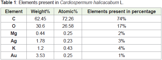
EDAX:
Energy Dispersive X-ray (EDX) spectrometer analysis confirmed
the presence of an elemental silver signal of the silver nanoparticles.
The number of X-ray counts are displayed on the vertical axis and
energy in KeV displayed on the horizontal axis. The silver (Ag) peak
is observed in the spectrum as well as in the elemental composition
showing its presence in the leaf extract (Table 1, Figure 3). Earlier,
Kumar et al. (2014) studied the characterization of AgNPs using XRD
and TEM analysis which showed a spherical shaped structure with an
average particle size of 25nm [10].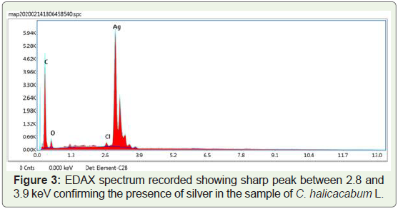
Nanoparticles act as ‘magic bullets’ that can target the desired
part of the plant to achieve their potential as herbicides, fungicides,
nutrients, fertilizers or nucleic acids [6]. Earlier, Farghaly and Nafady
(2015) used the leaf extract of Rosemary for the biosynthesis of
silver nanoparticles (AgNPs) and showed their eco-friendly and cost
effective nature. They have also studied the effect of AgNPs on the
growth of wheat and tomato plants [11-13]. Ashlesha et al. (2021) have
studied the green synthesis of nanoparticles and their antimicrobial
properties from ex-situ grown bryophytes. It uses agricultural inputs
more effectively and reduces the by-products that could harm the
environment as well as human health [14-17]. Logeswari et al. (2015)
studied the synthesis of nanoparticles from commercially available plant powders. Applications of Nanotechnology in agriculture can
prove to be a boon to mankind [18,19].
Conclusion
Silver nanoparticles find a large application in Industries and
medicine. Hazardous organic solvents and surfactants which are
often employed in chemical synthesis of nanoparticles can be
avoided through green synthesis techniques. The present study
demonstrates the bio-reduction of aqueous Ag+ ions by the leaf
extract of Cardiospermum halicacabum L. The present protocol is
an eco-friendly and cost-effective method for the synthesis of silver
nanoparticles. Further studies are to be carried out to analyze the
biological activities of the synthesized nanoparticles.
Acknowledgement
The authors thank the Bharat Ratna Prof. CNR Rao Research
Centre, Avinashilingam Institute for Home Science and Higher
Education for Women, Coimbatore for helping in the SEM and EDAX
analysis for the current study.
References
Citation
Hema J, Gayathri V, Sanandam J, Anitha R. Isolation and Characterization of Silver Nanoparticles in Cardiospermum halicacabum l. Leaf
Extract. J Plant Sci Res. 2022;9(2): 226