Research article
Sweet Potato (Ipomoea batatas L.): Suspension Culture for cell Aggregation
Md. Mahabubur Rahman1* and Rubaiyat Sharmin Sultana2
1Department of Crop Botany, Exim Bank Agricultural University Bangladesh, Chapainawabganj 6300, Bangladesh
2Department of Botany, University of Rajshahi, Rajshahi 6205, Bangladesh
Corresponding author: Md. Mahabubur Rahman, Lecturer, Department of Crop Botany, Exim Bank Agricultural University Bangladesh, Bangladesh; Tel: +88-01791499990; E-mail: spmahabub@yahoo.com
Citation: Rahman MM, Sultana RS. Sweet Potato (Ipomoea batatas L.): Suspension Culture for cell Aggregation. J Plant Sci Res. 2017; 4(2): 171.
Copyright © Rahman MM. 2017. This is an open access article distributed under the Creative Commons Attribution License,which permits unrestricted use, distribution, and reproduction in any medium, provided the original work is properly cited.
Journal of Plant Science & Research | ISSN: 2349-2805 | Volume: 4, Issue: 2
Submission: 14/08/2017; Accepted: 18/09/2017; Published: 06/10/2017
Abstract
Cell aggregation in suspension culture of sweet potato (Ipomoea batatas L.) was established from petiole-derived callus. The highest rate (55%) of petiole induced callus on agar-gelled Murashige and Skoog (MS) medium containing 10 μM 2,4-Dichlorophenoxyacetic acid (2,4-D) within 30 days of culture. These calli were used for preparation of suspension culture in liquid medium containing same concentration of 2,4-D that was used for callus induction. A growth curve with lag, exponential and stationary phages of cell growth was obtained when total amount of cell was plotted against the culture periods. Cells underwent divisions, resulting in free cells multiplication and cell aggregates formation after further 30 days of culture. The initial amount of cell during suspension culture showed effect in the cell proliferation, cell size and cell aggregate development. The additional amount of cell and cell aggregates formation were the highest when 5 ml Sedimented Cell Volume (SCV) of cells was cultured. The cell proliferation reduced and high number of elliptical cell was found in suspension when culture maintained for longer period without transferring to fresh medium.
Keywords:
Callus; Cell aggregate; Petioles; Sweet potato; Suspension culture
Introduction
Sweet potato (Ipomoea batatas L.) is one of the major food crops in the world. It is an important source of carbohydrates. Since sweet potato is a vegetatively propagated plant, the molecular breeding based genetic transformation is an important option to improve this plant. The successful genetic transformation cannot be achieved unless efficient plant regeneration protocol is established. Few reports were published on plant regeneration procedures of sweet potato through organogenesis and somatic embryogenesis on gelled medium. The ability of regeneration into plantlets using micropropagation system from apical meristems, lateral buds and nodal segments of sweet potato [1,2]. The callus induction and adventitious shoot formation procedure from storage root sections of sweet potato was established in vitro [3]. The clonal propagation of sweet potato through both adventitious and axillary procedures was established by Villordon and Labonte [4]. The embryogenic callus and efficient in vitro plant regeneration system were characterized by rapid and continuous production of somatic embryos [5-8]. The report on plant regeneration system using cell suspension culture has not been established, although one report on genetic transformation using somatic embryogenesis through cell suspension culture for sweet potato was published [9]. One report is insufficient for genetic transformation by somatic embryo genesis in suspension culture. Besides, lacking of detail information on somatic embryogenesis has remained in the protocol of Xing et al. [9]. Therefore, more detail research on cell proliferation, cell aggregate formation and their appearance in suspension culture of sweet potato is necessary for genetic transformation studies.
The aim of the present study is to establish efficient cell suspension culture for sweet potato. Herein, proliferation of cell and development of cell aggregates in suspension culture were studied in various media and culture conditions.
Materials and Methods
Basal medium preparation and culture conditions
MS basal medium was used for callus induction and cell suspension culture. All media were adjusted to pH 5.7 ± 0.1 and autoclaved at 121 °C for 20 min [10]. The medium was solidified with 0.8% (w/v) agar (Type M, Sigma), which was used for callus induction and the liquid medium was applied for cell suspension culture. The medium without Plant Growth Regulators (PGRs) received as control for all experiments. The cultures were maintained at 27 ± 1 °C under a 16-h photoperiod (35 μmol m-2 s-1).
Induction of callus and initiation of cell suspension culture
Leaves with petiole of field grown plant were collected between 10:00 to 11:00 am in the daytime with bright sunlight. Petioles were separated from leaves and washed thoroughly under running tap water for 30 min. Finally they were disinfected with 2% (v/v) solution of NaOCl for 10 min under laminar airflow followed by washing thrice with autoclaved distilled water. The disinfected petioles were sliced (approximately 1 cm in length) and cultured on MS medium containing either different concentrations of 2,4-Dichlorophenoxyacetic acid (2,4-D) at 5, 10 and 15 μM alone or in combinations with kinetin (KIN) at 0.5 μM.
Proliferated 30-day-old calli (1 g, fresh weight) on gelled MS medium containing 10 μM 2,4-D were cultured in 300 ml Erlenmeyer flask containing 50 ml liquid MS medium fortified with 10 μM 2,4- D. After 15 days of suspension culture, cells were proliferated in suspension cultures, which were used for further experiments.
Cell growth in batch culture
Cell proliferation in suspension culture was examined by batch culture. The amount of proliferated cells was determined by Sedimented Cell Volume (SCV) ml. For measuring of cell amount by SCV, suspension dispensed into sterile 100 ml measuring cylinders and allowed to sediment for 30 min. For batch culture, 5 ml SCV of cells from 15-day-old suspension culture were cultured initially in 300 ml Erlenmeyer flasks containing 50 ml MS liquid medium fortified with 10 μM 2,4-D. They were routinely transferred at one-weekinterval to a fresh medium and total amount (ml) SCV of cell per flask measured prior to transfer in each subculture. The suspension cultures were maintained up to 8 weeks.
Effect of initial cell amounts on cell proliferation, cell size and cell aggregates development
Cells from fifteen-day-old suspension were cultured initially at different volumes, 1, 3, 5, 10, 15, and 20 ml SCV in 300 ml Erlenmeyer flasks containing 50 ml of MS liquid medium supplemented with 10 μM 2,4-D. The cultures were transferred into fresh medium routinely in a week. After 4 weeks of culture, the proliferated cells in an Erlenmeyer flask were dispensed in a sterile 100 ml measuring cylinders and additional cell amount measured with SCV. In above all suspension culture, the number of cell aggregates was measured using hemacytometer. For obtaining the number of cell aggregates, 10 μl of liquid cultures were used. The number of cell aggregates was counted using the manual counter in each grid and the total number was calculated per 10 μl suspension and then total number of cell aggregates was counted from 1 ml suspension. For measurement of cell diameter from all suspensions, cells were observed under light microscope and photographs were taken. Radial and tangential diameters were measured by Image J software and average of radial and tangential diameters of each cell denoted as cell diameter.
Effect of subculture duration on cell size
Fifteen-day-old 5 ml SCV cells were cultured in MS containing 10 μM 2,4-D. Cell amount and appearances were observed in 6-weekold culture without transferring to the fresh medium and in 6-week old culture with regularly transferring at 7-day-interval. The types and size of cells were compared between these two conditions of cell suspension culture.
Statistical analysis
For callus induction, ten internode segments were cultured in one Petri dish, which was considered as one replication. To observe the effect of initial cell amount on the cell proliferation, cell size and cell aggregates number, four Erlenmeyer flasks were used and each flask was considered as one replication. From each flask, amount of cell (ml) SCV, diameters (twenty cells) and number of cell aggregates were measured. All experiments were repeated at least three times. The experiments were conducted with a completely randomized design and the data were analyzed by analysis of variance. The Least Significance Difference (LSD) test was used to distinguish differences among the mean value of treatments at 5% (p ≤ 0.05) level.
Results and Discussion
Callus induction
All variants of PGRs in MS medium were responsible for the callus induction except PGR-free MS medium. Of all PGR treatments examined, the callus induction frequency was the highest (55%) on MS medium containing 10 μM 2,4-D within 30 days of culture (Table 1). On this medium, callus induction was first observed 7 days after culture at the cut sides of petiole segments. After 30 days of culture, callus proliferation was surrounded throughout the explant. The induced callus color was pale-yellow (Figure 1). White calli alsoinduced on the rest of other PGRs with lower frequencies than that of10 μM 2,4-D. The significant differences (p ≤ 0.05) were observed inthe frequencies of explant inducing callus among the PGR treatmentsexamined here (Table 1). Alves et al. reported that the embryogenic callus was induced from lateral bud of sweet potato on MS medium fortified with 10 μM 2,4-D [7]. The medium containing 0.5-2.0 mg/l 2,4-D assisted to induce pale- to bright-yellow calli from leaf, shoottip, stem and root explants, which were responsible for embryos development [6]. Root discs of sweet potato cv. Leucorhiza produced callus and then developed meristemoids and shoots on various levels of Kn and 2,4-D combination [11]. From the above discussion, it is clarified that 2,4-D was essential for rapid callus induction, which is responsible for embryo or organ development of sweet potato. The trends of callus induction in the previous report is supported the present study, in which the best callus induction obtained from the petiole explants on medium containing 2,4-D alone and that could form proembryogenic aggregates.
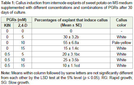
Table 1: Callus induction from internode explants of sweet potato on MS medium supplemented with different concentrations and combinations of PGRs after 30 days of culture.
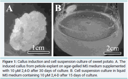
Figure 1: Callus induction and cell suspension culture of sweet potato. A. The induced callus from petiole explant on agar-gelled MS medium supplemented with 10 μM 2,4-D after 30 days of culture. B. Cell suspension culture in liquid MS medium containing 10 μM 2,4-D after 15 days of culture.
Cell growth by batch culture
The induced callus (1 g, fresh weight) on agar-gelled MS medium containing 10 μM 2,4-D were cultured in liquid MS medium containing 10 μM 2,4-D for 15 days (Figure 1). The cells of this suspension culture (5 ml SCV) were used for cell growth study in the batch culture. When the cell amount (SCV) is plotted against the culture period, a growth curve was obtained. The increase cell amount in the suspension culture was not observed from initial day to end of 2nd weeks of culture, indicating lag phage of cell growth. The cell growth gradually increased from 3rd to end of 7th week, which was indicated exponential growth phage. The subsequent one week (from 7th to end of 8th week) did not show cell growth, which is denoted as stationary phase (Figure 2). The biomass growth in the batch culture follows this growth carves with lag, exponential and stationary phase has been report by Razdan [12]. Krogstrup stated that lag phase was not clear in the growth curve but the cultures show a linear growth until day 26-28 and then cell proliferation eventually deteriorate in the cell suspension culture for Picea sitchensis [13].
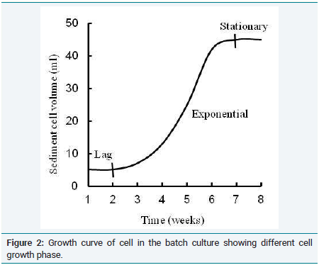
Figure 2: Growth curve of cell in the batch culture showing different cell growth phase.
Effects of cell initial cell amounts on cell proliferation, cell size and cell aggregates development
The initial amount of cell during suspension culture showed an important role in the cell proliferation, cell size and cell aggregate development in liquid MS medium containing 10 μM 2,4-D. The additional amount of cell (SCV) was gradually increased in the suspension culture with increasing initial amount of cell increased from 1 to 5 ml SCV and then it was gradually decreased when initial amount of cell from 10 to 20 ml (Table 2). The highest amount of additional cell was 8.2 ± 1.3 ml SCV, which was found in the suspension when initial cell amount was 5 ml SCV.
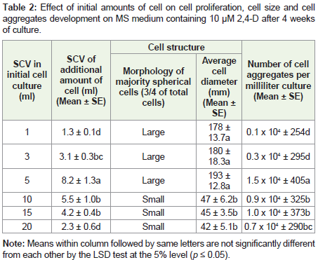
Table 2: Effect of initial amounts of cell on cell proliferation, cell size and cell aggregates development on MS medium containing 10 μM 2,4-D after 4 weeks of culture.
When initial amounts of cell were 1, 3 and 5 ml SCV in cell suspension cultures, majority free single cells (3/4 of total cells) in the suspension culture were large and spherical. The similar diameters of cell (178, 180 and 193 μm) were found in suspension when initial amounts of cell were 1, 3 and 5 ml SCV, respectively (Table 2). In rest of suspension cultures where initial amounts of cell were 10, 15 and 20 ml SCV, multiplied majority single cells (3/4 of total cells) were small and spherical, and the diameters of cell were similar 47, 45 and 42 μm, respectively. The diameter of cell differed significantly in the amount of cell during initiation of culture (Table 2).
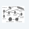
Figure 3: A diagrammatic figure of cell division and cell aggregates formation in the suspension culture.
After 30 days of culture, free cells and cell aggregates were observed in cell suspension culture. In the suspension culture, cells underwent mitosis divisions, resulting in free cells multiplication and early proembryogenic structure like two-, three-, four-, octa-, 16-celled early proembryogenic structure and multicellular aggregate (proembryo) formation (Figure 3). The filamentous four-celledembryogenic structure was also found in this suspension culture (Figure 3). In this experiment, two-celled, three-celled, four-celled early proembryos and Multicellular aggregates (proembryos) were observed (Figures not shown). The number of cell aggregates was gradually increased when initial cell amount was increased from 1 to 5 ml SCV. Decrease in the number of cell aggregates was found when initial cell amount was increased from 10 to 20 ml SCV in liquid medium (Table 2). The initial cell amount used in a given culture system can have dramatic effects on cell proliferation [14]. The cell size and cell aggregate formation in the cell suspension culture was influenced by the initial cell amount [15].
Effect of subculture duration on cell size
For long time maintenance of cell suspension culture, polyphenols are accumulated in the liquid medium, which restricted cell division and cell enlargement [16]. In the present study, cell amount was lower (32 ml SCV) in a medium for long time (6 weeks) culture without subculturing to fresh medium than regular subculture to fresh MS medium (45 ml SCV) (Table 3). The reduction of cell amount in suspension without subculturing to fresh medium might be occurred with the extensive accumulation of polyphenols. When cell culture was maintained for a long time (6 weeks) without subculturing to fresh MS medium containing 10 μM 2,4-D, the most of the cells shape was elliptical and large-sized (range 50-210 μm in diameter) (Figure 4). The spherical cells were also observed in this culture condition but they were smaller size (range 30-100 μm in diameter) than that of elliptical cells. In this culture, elliptical cell number was higher than spherical cell (elliptical cell: spherical cell was 1: 0.35). On the other hand, when cell culture regularly transferred to fresh medium at 7-day-intervel, the number of spherical cell was higher than elliptical cell (elliptical cell: spherical cell was 1: 7) (Table 3). The diameters of elliptical cells (range 40 - 150 μm in diameter) were higher than spherical cells (range 30 - 80 μm in diameter) (Figure 4). In the present experiment, the higher number of elliptical cells in 6-week-old culture without subculturing may form at the presence of PGR. The contrast trend was reported by Häsler, who observed that elliptical and spherical cells formed in ten-year-old sugar beet leaf-derived callus when callus was maintained on agar-gelled MS medium supplemented with 0.1 mgl-1 2,4-D and 0.1 mgl-1 BAP, while only spherical cells were found in ten-year-old callus gown on PGRfree MS medium.
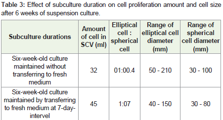
Table 3: Effect of subculture duration on cell proliferation amount and cell size after 6 weeks of suspension culture.
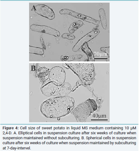
Figure 4: Cell size of sweet potato in liquid MS medium containing 10 μM 2,4-D. A. Elliptical cells in suspension culture after six weeks of culture when suspension maintained without subculturing. B. Spherical cells in suspension culture after six weeks of culture when suspension maintained by subculturing at 7-day-intervel.
Conclusion
The present study established cell suspension culture of sweet potato. For establishment of more efficient plant regeneration procedure in comparison with previously established protocol by Xing et al., the knowledge in cell suspension culture established here will be cooperative [9]. The present study will also be helpful to establish single cell origin of plant as well as suitable genetic modification processes for sweet potato.
References
- Litz RE, Conover RA (1978) In vitro propagation of sweet potato. Hort Sci 13: 659-660.
- De Castro OF, De Andrade AG (1995) Meristem culture of sweet potato (Ipomoea batatas L.). Braz Agricu Res 30: 917-922.
- Hwang LS, Skirvin RM, Casyao J, Bouwkamp J (1983) Adventitious shoot formation from sections of sweet potato grown in vitro. Sci Hort 20: 119-129.
- Villordon AQ, Labonte DR (1996) Genetic variation among sweet potatoes propagated through nodal and adventitious sprouts. J Am Soc Hort Sci 121: 170-174.
- Zheng Q, Dessai AP, Prakash CS (1996) Rapid and repetitive plant regeneration in sweet potato via embryogenesis. Plant Cell Rep 15: 381-385.
- Liu JR, Cantliffe DJ (1984) Somatic embryogenesis and plant regeneration in tissue culture of sweet potato ((Ipomoea batatas Poir.). Plant Cell Rep 3: 112-115.
- Alves JM, Sihachakr D, Allot M, Tizroutine S, Mussio I, et al. (1994) Isozyme modifications and plant regeneration through somatic embryogenesis in sweet potato (Ipomoea batatas (L.) Lam.). Plant Cell Rep 13: 437-441.
- Chee RP, Cantliffe DJ (1988) Somatic embryony patterns and plant regeneration in Ipomoea batatas Poir. In Vitro Cellular Develop Biol 24: 955-958.
- Xing YJ, Ji1 Q, Yang Q, Luo YM, Li Q, et al. (2008) Studies on Agrobacterium-mediated genetic transformation of embryogenic suspension cultures of sweet potato. Afr J Biotech 7: 534-540.
- Murashige T, Skoog F (1962) A revised medium for rapid growth and bioassays with tobacco tissue cultures. Plant Physiol 15: 473-497.
- Aloufa MA (2002) Some factors affecting the callus induction and shoot formation in two cultivars of sweet potato (Ipomoea batatas (L.) Pois). Sci Agrotechnol 26: 964-969.
- Razdan MK (1993) An introduction to plant tissue culture, (2nd edn). Science Publishers, Delhi, India. pp. 1-367.
- Krogstrup P (1990) Effect of culture densities on cell proliferation and regeneration from embryogenic cell suspensions of Picea sitchensis. Plant Sci 72: 115-123.
- Bhojwani SS, Razdan MK (1996) Cell culture. In: Bhojwani SS, Razdan MK (Eds), Plant tissue culture: Theory and practice, a revised edition. Elsevier, Amsterdam, New York. pp. 63-93.
- Kubota C, Chun C (2003) Transplant production in the 21st century: Proceedings of the international symposium on transplant production in closed system for solving the global issues on environmental conservation, food, resources and energy. Springer Science & Business Media, The Netherlands, pp. 1-290.
- Davies ME (1972) Polyphenol synthesis in cell suspension cultures of Paul's Scarlet rose. Planta 104: 50-65.
- Häsler J, Wüest J, Gaspar T, Crèvecoeur M (2003) A long term in vitro-cultured plant cells show typical neoplastic features at the cytological level. Biol Cell 95: 357-364.