Study of Phytochemicals, Trace Elements and Antibacterial Activity of Silybum marianum (L.)Gaertn.
Shweta Puri1, M C Sidhu1*, Rupinder Tewari2 and Avantika Sharma2
Corresponding author: Dr. M C Sidhu, Department of Botany, Panjab University, Chandigarh, India; E-mail: mcsidhu@gmail.com
1Department of Botany, Panjab University, Chandigarh, India
2Department of Microbial Biotechnology, Panjab University, Chandigarh, India
Citation: Puri S, Sidhu MC, Tewari R, Sharma A. Study of Phytochemicals, Trace Elements and Antibacterial Activity of Silybum marianum (L.) Gaertn.J Plant Sci Res. 2015;2(2): 122.
Copyright © 2015 M C Sidhu et al. This is an open access article distributed under the Creative Commons Attribution License, which permits unrestricted use, distribution, and reproduction in any medium, provided the original work is properly cited.
Journal of Plant Science & Research | ISSN: 2349-2805 | Volume: 2, Issue: 1
Submission: 20/12/2014; Accepted: 16/01/2015; Published: 21/02/2015
Abstract
Silybum marianum is a biennial herbaceous plant of family Asteraceae. It is used traditionally to treat both human and animal ailments in different parts of the world. Keeping in view the medicinal importance of this species, it has been selected for the present study. The phytochemical analysis has revealed the presence of various phytoconstituents like alkaloids, amino acids, carbohydrates, flavonoids, glycosides, gums and mucilage, phenolics, reducing sugars, tannins etc. The Fourier Transform Infrared (FTIR) spectroscopy has provided the details of various functional groups including amides, esters, aldehydes etc. with different peak values associated with the screened phytochemicals. The Wavelength Dispersive X-Ray Fluorescence (WD-XRF) analysis has shown the presence of elements like Potassium, Calcium, Chlorine etc. Phytochemicals and other elements are likely to be responsible for the medicinal potential of this species. It has also shown antibacterial activity against some bacterial strains.
Keywords: Phytochemicals; Antibacterial activity; FTIR spectroscopy; WD-XRF analysis
Abbreviations: Aq: Aqueous; Eth: Ethanol
Introduction
Silybum marianum L. Gaertn. (milk thistle) is a member of family Asteraceae. It is a native of Southern and Western Europe, South America and North America in the Eastern United States and California [1]. It is five to ten feet in height, biennial herb. Leaves are hard, green, shiny and have spiny edges and white veins. Reddish purple flower is solitary, borne at the tip of the branch with sharp spiny bracts. The seeds are hard and have white silky pappus [1,2].
This plant has a potential antioxidant activity [3] along with anticancer, antidepressant, cardio protective, hepatoprotective, immunostimulative and neuroprotective activity [4]. It works as antidote against Amanita mushroom poisoning [5]. Ripe seeds are used in the treatment of kidney and various hepatobiliary problems, such as hepatitis, cirrhosis, gallstones and jaundice [6]. This plant also plays an important role in endocrine function as antidiabetic and pancreatic protectant [7]. It contains phytoconstituents like flavonoids, phenols and tannins [8], amino acids, saponins, sugars [9], flavonoids [10], alkaloids, carbohydrates, steroids and tannins [11]. Antibacterial activity of seeds has been reported against Bacillus subtilis, Escherichia coli, Pseudomonas aeruginosa, Salmonella typhi etc. [12]. The chloroform and ethanol extracts of S. marianum leaves and hypocotyls callus have shown antibacterial activity against different Gram +ve and Gram –ve bacteria [9]. Therefore, the present study was undertaken to evaluate the phytochemicals, FTIR spectroscopy and WD-XRF analysis.
Materials and Methods
Plant materials
Silybum marianum has been selected for the present study. The plant material was collected from the Panjab University, Chandigarh campus during 2012-2013. The collected material was washed thoroughly with tap water and then with distilled water. Plant specimen was pressed and dried to prepare the herbarium sheet. The voucher specimen has been deposited in the Herbarium, Department of Botany (PAN No.-20331), Panjab University, Chandigarh, India.
Preparation of extracts
The collected plant material was washed thoroughly and allowed to dry at room temperature. The dried material (whole plant) was powdered by using an electric grinder. Aqueous and ethanol extracts of Silybum marianum has been prepared by using the following methods:
Preparation of aqueous extract
20gm of plant powder was added to 100ml of distilled water in a conical flask. The mixture was kept on an orbital shaker for 24 hours at room temperature. Then it was filtered through folds of muslin cloth. Further, it was again filtered through Whatmann’s Filter No. 1 and the filtrate was collected in vials. The final extract obtained is a stock solution of 200mg/ml concentration. The extract was stored in refrigerator at 4°C till further use [13].
Preparation of ethanol extract
The ethanol extract was prepared using the Soxhlet extraction method [14]. Powdered plant material (10gm) was extracted with ethanol (130ml) in a Soxhlet apparatus. The extract was allowed to evaporate at room temperature till it reaches the 1/3rd volume of the original extract. Then it was stored at 4°C in vials.
Phytochemical screening
Phytochemical analysis of both aqueous and ethanol extracts were carried out for the presence of various phytoconstituents such as alkaloids [15], carbohydrates [16,17], flavonoids [15,16], glycosides [16,18], gums and mucilage [16,18], phenols [16,18], phlobatannins [19], reducing sugars [20], saponins [15], steroids [15], tannins [18,20] and terpenoids [21] using standard procedures.
Bacterial strains
The antibacterial activity of Silybum marianum extracts against four bacteria namely Enterococcus faecalis (MTCC 439), Staphylococcus aureus (ATCC 25923) (Gram +ve), Escherichia coli(ATCC 25922) and Pseudomonas aeruginosa (ATCC 27853) (Gram –ve) has been studied.
Preparation of inoculum
A single colony of bacterial strain was inoculated from a streak plate into 10ml Mueller Hinton Broth media [g/L: beef infusion 300, casein acid hydrolysate 17.5, starch 1.5 and pH 7.4 ± 0.1] containing tubes. These tubes were incubated at 37°C/200rpm for 16-18 hours. This is a primary culture. 100μl of primary culture was inoculated into 10ml MHB media. The tubes were incubated at 37°C/200rpm till absorbance reaches 0.3 at 600nm.
Antibacterial activity using agar well diffusion method
100μl of culture was spread on MHA (Mueller Hinton Agar) plates and allowed to dry. Wells were made with the help of a sterile cork borer. The dose of 20μl, 50μl and 100μl was used initially. But these doses have not shown any zone of inhibition after 24 hours. Thus the volume of the dose was increased to 150μl and 200μl. Different doses of plant extracts i.e., 150μl and 200μl were added to the wells along with aqueous and organic solvent as control. These plates were incubated at 37°C overnight. The time interval of 24 hours was selected as the activity of extracts remained same even after 48 hours. But contamination in some cases has been observed near 48 hours time interval. To avoid contamination, the present time interval was selected. After incubation, the plates were observed for zone of inhibition and the diameter of zone was measured.
Fourier Transform Infrared (FTIR) Spectroscopy has been done using Perkin Elmer Spectrum 400 FT-IR/ FT-FIR spectrometer. Based on IR spectra various functional groups were detected in plant powder of this species. This test has been carried out to authenticate the preliminary phytochemical observations. The Wavelength Dispersive X-Ray Fluorescence (WD-XRF) analysis was done to study the presence of micro and macro elements.
Results and Discussion
Phytochemical screening
The different phytochemicals like alkaloids, carbohydrates, flavonoids, glycosides, gums and mucilage, phenolics, phlobotannins, reducing sugars, saponins, steroids, tannins and terpenoids have been reported during the present investigation. Both aqueous and ethanol extracts of Silybum marianum, revealed the presence of many phytoconstituents. All the studied phytoconstituents except alkaloids, reducing sugars and saponins were present in aqueous extract. Presently studied ethanol extract contains all the phytoconstituents under consideration except saponins (Table 1). Shah et al. [8] observed flavonoids, phenols and tannins during phytochemical evaluation of aqueous extract this plant. John and Koperuncholan [9] analyzed the ethanol extracts of leaf and hypocotyls callus. They have reported the presence of amino acids, sugars and tannins. Alkaloids, flavonoids, phenolics, saponins, steroids etc. were absent in their study. The difference in phytochemical observations is likely due to the use of different plant parts. The details of the phytochemicals screened during the present study are shown in Table 1.
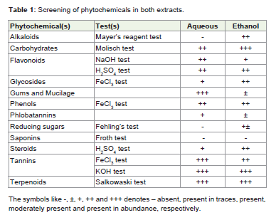
Table 1: Screening of phytochemicals in both extracts.
Antibacterial activity
The extracts of Silybum marianum have been screened for antibacterial activity against two Gram +ve (Enterococcus faecalis, Staphylococcus aureus) and two Gram –ve (Escherichia coli, Pseudomonas aeruginosa) bacterial strains. Different doses of both aqueous and ethanol extracts (150μl, 200μl) were given to all the bacterial strains under investigation. Extracts were tested along with ethanol and distilled water (DW) as a control. A 150μl dose of aqueous extract formed a 5mm inhibition zone against E. faecalis. But the similar dose of ethanol extract was found active against E. coli only with a 2mm zone of inhibition. At a higher dose (200μl), the ethanol extract have shown activity against all the four bacterial strains under investigation with 11mm, 7mm, 5mm, and 2mm zones of inhibitions for S. aureus (ATCC 25923), P. aeruginosa ( ATCC 27853), E. coli (ATCC 25922) and E. faecalis (MTCC 439), respectively (Figures 1-6; Table 2). Antibacterial activity of aqueous and ethanol extracts of S. marianum seeds was studied by Hassan et al. [12] against Bacillus subtilis, Escherichia coli, Klebsiella pneumonia, Pseudomonas aeruginosa, Proteus vulgaris, Staphylococcus aureus and Salmonella typhi with methicillin and oxacillin antibiotic as a control. Extracts were found active against all bacteria. Their study has supported the present findings. Similarly, the silymarin isolated from the ethanol extract of S. marianum had shown activity against B. subtilis (NCTC 8236), P. vulgaris (ATCC 6380), S. aureus (NCTC 25953) with 17mm, 15mm and 21mm inhibition zones, respectively. This extract was found active only against Gram +ve bacteria However, during the present study, ethanol [8]. However, during the present study, ethanol extract have shown activity against Gram +ve and Gram –ve bacterial strains. This difference in activity of the extracts is likely due to the use of higher dose (200μl). The phytochemicals like alkaloids and flavonoids along with micro and macro elements are likely to be responsible for the antibacterial activity of this species.
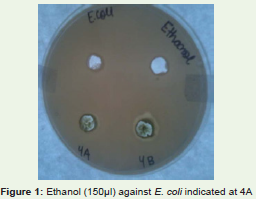
Figure 1: Ethanol (150μl) against E. coli indicated at 4A
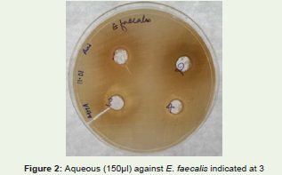
Figure 2: Aqueous (150μl) against E. faecalis indicated at 3
Figure 3: Ethanol (200μl) against E. coli indicated at 4B
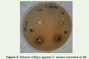
Figure 4: Ethanol (200μl) against S. aureus indicated at 4B
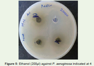
Figure 5: Ethanol (200μl) against P. aeruginosa indicated at 4
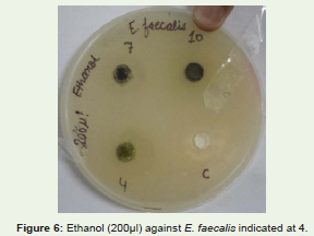
Figure 6: Ethanol (200μl) against E. faecalis indicated at 4.
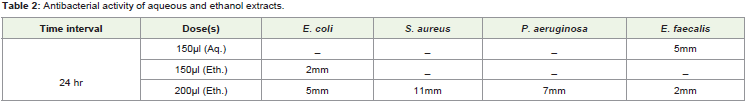
Table 2: Antibacterial activity of aqueous and ethanol extracts.
FTIR absorption spectra of whole plant powder have been studied. The observed IR frequencies pertaining to different functionalities like 3271cm-1 (N-H stretch in amines and amides); 2921cm-1(alkanes sp3 C-H stretch); 2221cm-1, 2166cm-1, 2033cm-1, 1997 (CC stretch, CN stretch, -N=C=O stretch, -N=C=S stretch, -C=C=C stretch in allenes and –C=C=O stretch in ketenes); 1734cm-1 (C=O stretch in amides, esters, acids, aldehydes and ketones); 1587cm-1 (N-H bend in amides and nitro group absorption); 1377cm-1 (-CH3 bend in alkanes and nitro group absorption); 1319cm-1 (C-N stretch in amines, C-O stretch in carboxylic acids, asymmetric S=O stretch in sulphones, sulphonyl chlorides, sulphates and sulphonamides); 1241cm-1 (C=O bending in ketones, phenyl alkyl ethers, C-O stretch in alcohols, ethers, esters, carboxylic acids, anhydrides, C-N stretch in amines and aryl fluorides); 1160cm-1, 1030cm-1(C-N stretch in amines, C-O stretch in alcohols, ethers, esters and S=O symmetric stretch sulphonates); 669cm-1 (alkenes out of plane bonds and C-Cl stretch in aliphatic chlorides) and 558cm-1, 443cm-1, 408cm-1(C-X for bromides/ iodide).
The IR absorption frequency values are corresponding to different functional groups of various phytochemicals (Figure 7). The WDXRF analysis of whole plant powder has provided qualitative and quantitative details of the micro and macro elements present (Table 3). These are known for their medicinal potentiality for the treatment of different ailments. Thus, further studies are required to establish their role.
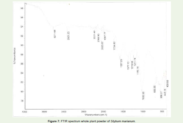
Figure 7: FTIR spectrum whole plant powder of Silybum marianum.
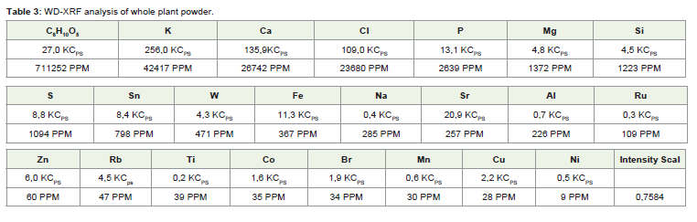
Table 3: WD-XRF analysis of whole plant powder.
Conclusion
The phytochemical screening of aqueous and ethanol extracts of this plant have revealed the presence of alkaloids, carbohydrates, flavonoids, glycosides, gums and mucilage, phenolics, phlobotannins, reducing sugars, steroids, tannins and terpenoids. The FTIR spectroscopy of whole plant powder has shown the presence of various functional groups such as amines & amides, alkanes, esters, acids, aldehydes and ketones etc. WD-XRF analysis has revealed the presence of various elements like K, Ca, Cl, Mg, Si, S etc. A similar dose of (150μl) of both the extracts has shown activity against different bacteria. The increased dose (200μl) was found active against all the four bacterial strains. Some of the phytochemicals and elements are likely to be responsible for the antibacterial activity. The analysis of several populations of this species may give rise to different chemotypes having comparative amount of the chemicals of interest. Further study can be performed to isolate the different phytoconstituents and to see their individual activity against bacterial strains. Similarly, the role of different elements can also be studied in detail after separating them using standard procedures.
Acknowledgement
Authors are grateful to Professor A. S. Ahluwalia, Chairperson, Department of Botany, Panjab University, Chandigarh for providing necessary facilities during the present investigation.
References
- Murphy JM, Caban M, Kemper KJ (2000) Milk thistle (Silybum marianum). Longwood Herb Task Force Center Holistic Pediatr Ed Res 1-25.
- Sidhu MC, Saini P (2012) Silybum marianum: a plant of high medicinal importance- a review. World J Pharm Res 1: 72-86.
- Mira ML, Azevedo MS, Manso C (1987) The neutralization of hydroxyl radical by silibin, sorbinil and bendazac. Free Radic Res Commun 4: 125-129.
- Post-White J, Ladas EJ, Kelly KM (2007) Advances in the use of Milk thistle (Silybum marianum). Integr Cancer Ther 6: 104-109.
- Flora K, Hahn M, Rosen H, Benner K (1998) Milk thistle (Silybum marianum) for the therapy of liver disease. Am J Gastroenterol 93: 139-143.
- Fintelmann V (1997) Modern phytotherapy and its uses in gastrointestinal conditions. Planta Med 57: S48-52.
- Velussi M, Cernigoi AM, De Monte A, Dapas F, Caffau C, et al (1997) Long- term (12 months) treatment with an anti-oxidant drug (silymarin) is effective on hyperinsulinemia, exogenous insulin need and malondialdehyde levels in cirrhotic diabetic patients. J Hepatol 26: 871-879.
- Shah SMM, Khan FA, Shah SMdH, Chishti, KA, Pirzada, et al. (2011) Evaluation of phytochemicals and antimicrobial activity of white and blue capitulum and whole plant of Silybum marianum. World Appl Sci J 12: 1139-1144.
- John SA, Koperuncholan M (2012) Direct root regeneration and indirect organogenesis in Silybum marianum and preliminary phytochemical, antibacterial studies of its callus. Int J Pharm 2: 392-400.
- Lahlah ZF, Meziani M, Maza A (2012) Silymarin natural antimicrobial agent extracted from Silybum marianum. J Acad 2: 164-169.
- Balian S, Ahmad, S, Zafar R (2006) Anti-inflammatory activity of leaf and leaf callus of Silybum marianum (L.) Gaertn. in albino rats. Indian J Pharm 38: 213-214.
- Hassan A, Rahman S, Deeba F, Mahmud S (2008) Antimicrobial activity of some plant extracts having hepatoprotective effects. J Med Plants Res 3: 20-23.
- Gupta VP, Govindaiah, Datta RK (1996) Plant extracts: a non-chemical approach to control Fusarium disease of mulberry. Curr Sci 71: 406-409.
- Yadav RNS, Agarwala M (2011) Phytochemical analysis of some medicinal plants. J Phytol 3: 10-14.
- Idu M, Igeleke CL (2012) Antimicrobial activity and phytochemistry of Khaya senegalensis roots. Int J Ayur Herb Med 2: 415-422.
- Trease GE, Evans WC (1989) Trease and Evans pharmacognosy. (13thedn) ELBS, London.
- Kokate CK (1994) Practical pharmacognosy. Vallabh Prakashan, Delhi, India.
- Sofowora A (1993) Medicinal plants and traditional medicine in Africa. Spectrum Books Ltd., Ibadan.
- Ajayi IA, Ajibade O, Oderinde RA (2011) Preliminary phytochemical analysis of some medicinal plant seeds. Res J Chem Sci 1: 58-62.
- Evans WC (2002) Trease and Evans pharmacognosy. (15thedn), W.B. Saunders and Company, London.
- Harborne JB (1998) Phytochemical methods: A guide to modern techniques of plant analysis. Chapman & Hall Std., London, New York.