


Research Article
Anatomical and Pharmacognostic Study of Leaf and Stem of a Medicinally Important Plant, Senna sophera (L.) Roxb. in and Around Santiniketan, West Bengal
Ghosh R, Mandal S and Roy A*
Department of Botany, Cytology and Plant Tissue Culture Laboratory, Visva- Bharati, India.
*Corresponding author:Dr. Anjalika Roy, Department of Botany, Cytology and Plant Tissue Culture Laboratory, Visva- Bharati, India. E-Mail Id: anjalika.roy@visva-bharati.ac.in
Copyright: © Ghosh R, et al. 2026. This is an open access article distributed under the Creative Commons Attribution License, which permits unrestricted use, distribution, and reproduction in any medium, provided the original work is properly cited.
Article Information:Submission: 06/07/2026; Accepted: 29/07/2026; Published: 31/07/2026
Abstract
The current research focuses on the anatomical and pharmacognostic profiles of the leaves and stem of Senna sophera (L.) Roxb., belonging to the family Fabaceae (subfamily Caesalpinioideae). This plant is a widely distributed shrub found in tropical and subtropical regions, especially across Asia and Africa. In India, especially in West Bengal, this plant is found on roadsides and in wasteland areas. Traditionally, the leaf and stem of this plant have
been used for treating health conditions such as cough, fever, rheumatism, and skin diseases. The leaf micromorphology study shows the wavy outline of epidermal cell walls with irregularly shaped cells on both the adaxial and abaxial surfaces. Notably, the epidermal cells on the adaxial surface (35.1±1.2μm x
29.31±1.1μm) were larger than those on the abaxial surface (28.13±1.2μm x 12.9±1.1μm). Paracytic stomata were present on both surfaces of the leaf. The stomatal index values were 16±1.4 and 21±1.8 on the upper and lower surfaces, respectively. Unicellular non-glandular trichomes were observed in both stem and leaves. The trichome index of the upper epidermis was 2.31. The moisture content and total Ash value of leaves were estimated to be 10.66% and 6.64%, respectively. Microchemical tests confirmed the presence of alkaloids, steroids, saponins and flavonoids. Most of these findings could provide the scientific criteria for correct identification and establishment of the standard of both drug parts of this plant. Additional research is necessary to evaluate the potential of phenolic compounds as an alternative to pharmacological treatments.
Keywords:Senna sophera L; Foliar micromorphology; anatomy; xylem element; microchemical study.
Introduction
Herbal plants have been documented in the Ayurvedic and
Siddha systems of medicine, which play a vital role in traditional
health care for human beings since ancient times. Their utilization
as natural therapeutic agents for various ailments are well
documented across numerous indigenous societies (Begum and
Rahaman, 2021) [1]. The Traditional Medicine Feature provides
insights into worldwide ancient practices that contribute to
conventional medicines (Ghawte et al., 2025) [2]. According to the
World Health Organization (WHO), a substantial proportion of
the global population relies on plant-based medicines, with most
traditional therapies employing either whole plant extracts or their
active constituents (Begum and Rahaman, 2021) [1]. Indigenous
knowledge systems document millennia of plant, animal, and
mineral use for health care. This oral wisdom forms the foundation
of codified traditional medicines and modern pharmacology,
providing locally accessible remedies for everything from common
colds to chronic ailments across global societies (Alum, 2024).
This voluminous knowledge of medicinal plants for health care
since the inception of civilization is a good source of raw materials
for modern medicine nowadays; however, adulteration remains
a major concern, often involving the substitution or addition of
substandard materials. Such issues necessitate the implementation
of robust quality control protocols to ensure the purity and
efficacy of crude herbal drugs. In drug discovery and development,
knowledge of the traditional uses of crude plant products plays a
significant role (Sultana et al., 2023; Garzon-Castano et al., 2018) [3,4].
Standardizing and documenting the characteristics of raw materials
used in the preparation of herbal medicines is of utmost importance
to verify the authenticity of the herbal raw ingredients (Bhadury
et al., 2025) [5]. Reliable identification and evaluation of herbal
drugs can be achieved through pharmacognostic investigations,
encompassing morpho-anatomical characterization, organoleptic
assessment, physicochemical profiling, and both preliminary and
advanced phytochemical screening.
The genus “Senna” belongs to the family Fabaceae, subfamily
Caesalpinioideae, and is an economically important flowering plant.
Bark and oil extracts of Senna species are used for flavouring, soap,
candy, and perfumery (Rahman et al., 2013) [6]. Senna sophera
L. (Roxb.) is a widely distributed shrub found in tropical and
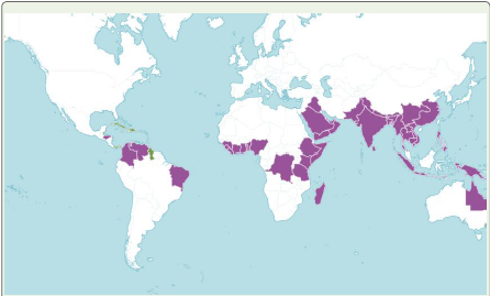
subtropical regions, especially across Asia and Africa and is also
native to the Indian subcontinent and Africa. In India, this plant
is found in forests, wasteland areas, and roadside areas of Andhra
Pradesh, Karnataka, Odisha, Tamil Nadu, and West Bengal. The
plant is known by its vernacular name in various parts of India,
viz. Kasondi (Hindi), Kasamarda (Sanskrit), Junglitakla (Marathi),
Ghodachakumda (Oriya), Kalkasunda (Bengali), Ponnavarai (Tamil),
Thounam (Manipuri), etc. (Ghosh et al., 2025) [2]. Traditionally,
various parts of S. sophera L.(Roxb.), including leaves, roots,
and seeds, have been used to treat ailments such as cough, fever,
rheumatism, and skin diseases (Chopra et al., 1956; Nadkarni,
1976).
It is reported that the plant Senna sophera is used in
Ayurveda, Unani and folk medicine in the treatment of asthma,
bronchitis, allergic rhinitis, ringworm infection, skin infection,
diabetes, piles, jaundice, fever, rheumatoid arthritis, joint pain,
gastrointestinal diseases, and epilepsy (Ghosh et al., 2025; Bilal et
al., 2005) [2]. Phytochemical investigations have revealed the
presence of flavonoids, anthraquinones, glycosides, tannins, and
saponins, which are believed to be responsible for its diverse
pharmacological activities (Harborne, 1998; Khandelwal, 2008).
Saha et al., 2012 and Mondal et al., 2015 [7] have demonstrated
the plant’s antioxidant, antimicrobial, hepatoprotective, and
anti-inflammatory properties, making it a promising candidate
for therapeutic applications. Despite the existing research on
the pharmacology and phytochemistry of Senna sophera, there are
still gaps in the comprehensive evaluation of the pharmacognostic
attributes of its leaf and bark. No previous studies have specifically
examined their pharmacognostic characters. Therefore, the main
objective of this study was to investigate the anatomical and
pharmacognostic properties of both the leaf and stem of Senna
sophera, as these parts are commonly used as crude drugs for various
medicinal purposes.
Materials And Methods
Material:
Scientific Name: Senna sophera (L.) Roxb., annual or biennial
undershrub.(https:/ /powo. s c i enc e .kew.or g / t a xon/urn: l s id: ipni . org:names:518364-1)
Study area: The different places of Santiniketan and Sriniketan were chosen as the study area. UNESCO has declared Santiniketan as a World Heritage Site surrounded by lush greenery. However, an increasing number of tourists, private vehicles and dust are the major pollution threats in Santiniketan.
Place of work and collection time: Anatomical and pharmacognostic studies were carried out in the Cytology and Plant Tissue Culture Laboratory during the year 2025. The plant material was collected during March -June and August- November in the year of 2025.
Methods:
Plant identification and herbarium preparation: Fresh twigs
were collected from a mature plant grown near Kali Shayer Temple,
Canal Road, Sriniketan (23° 40’ 10.038” N 87° 40’ 5.8836” E). The
plant has been identified with the help of available literature (Sanyal,
1994) [8] and identification was confirmed through consultation
with an expert taxonomist. The nomenclature of the species has been
updated following the standard website, like ‘Plants of the World
Online’ (http://powo.science.kew.org/). After collection, the plant
specimen has been processed, and the herbarium was prepared
following the techniques suggested by Jain and Rao (1977). For future
reference, the herbarium specimen was submitted to CAL.[Voucher specimen number: VB/RG-1C].
Leaf extract preparation of plant: Collected leaf samples of S.
sophera were washed thoroughly, shade-dried, and ground into
fine powder. The leaf powder was stored in an airtight vessel at 4 °C
for future use. The 10 gm of powder sample of each plant part was
extracted with 150 mL of 80% aqueous methanol in a 250 mL conical
flask, kept in a mechanical shaker at 28±2°C for 36 h.28 ± 2 °C for
24 h, and the same extraction process was repeated three times. The
methanolic extract of the leaf was filtered with Whatman’s No.1 filter
paper. The filtrate was subjected to evaporation at room temperature
(28 ± 2 °C). The ultimate extract yield was stored at 4 °C and dissolved
in dimethyl sulfoxide (DMSO) to make the stock solution of extract
before use.
Study of foliar micromorphology: Leaf samples were cleared off
the chlorophylls following Bokhari’s method (Bokhari, 1970). The
cleared leaf samples were then mounted on the slide with a drop of
10% glycerine and 1% safranin and observed under compound light
microscope (ZEISS, AXIOSTAR plus, model number 176045). For
field emission scanning electron microscope (FE-SEM) analysis, the
leaf specimens were prepared following the methodology of Yuan
et al. (2020), and subsequently examined under a FE-SEM (Gemini
SEM 450; Serial No. 8216010130), from which suitable micrographs
were captured.
Vegetative anatomy-An anatomical study was done by cutting
freehand sections of freshly collected leaf, petiole, and wood of the
selected plant. All stained sections (Staining followed by the double staining
method {Johansen, 1940}) were subsequently observed
under a compound light microscope (ZEISS, AXIOSTAR plus, model
number 176045).

Xylem element study- Thin wooden pieces, approximately 1
cm in length, are boiled in 10%–40 % nitric acid (HNO₃) for 10–12
minutes. After decanting the acid, the sample is again boiled in 10%–
40 % potassium hydroxide (KOH) solution for another 10-12 min.
The KOH solution is then decanted, and the sample is washed 2-3
times with tap water. A small portion of the boiled wood sample is
placed on a glass slide, teased apart using dissecting needles, stained
with 1% safranin, and mounted in 10% glycerin (WHO, 1998) [9].
The prepared slide is then observed under a microscope (ZEISS,
AXIOSTAR plus, model number 176045).
Organoleptic study- The study was conducted on the powdered leaf sample with the help of sensory organs. This involved assessing various properties, including colour, odour, taste and texture of the crude drugs (WHO, 1998) [9].
Physicochemical evaluation-The physicochemical characteristics of the leaf powder were evaluated (Evans, 2008) [10]. This evaluation included the determination of moisture content and, ash value (total ash, acid-insoluble ash, and water-soluble ash) of the plant samples.
Organoleptic study- The study was conducted on the powdered leaf sample with the help of sensory organs. This involved assessing various properties, including colour, odour, taste and texture of the crude drugs (WHO, 1998) [9].
Physicochemical evaluation-The physicochemical characteristics of the leaf powder were evaluated (Evans, 2008) [10]. This evaluation included the determination of moisture content and, ash value (total ash, acid-insoluble ash, and water-soluble ash) of the plant samples.
Preliminary microchemical colour reaction tests of leaf and
stem powder: The methanolic extract of leaf and bark powder was
obtained by cold maceration technique. The extracts were then
screened for the detection of different phytochemical groups by
chemical colour reaction tests following standard methods (Bush and
Taylor, 1952; Evans, 2009) [10].
Statistical Analysis
One-way analysis of variance was performed on all data (ANOVA) given as the mean±standard deviation of three copies. Difference among the average values of all the resulting data was compared using Tukey’s HSD test (honestly significant difference; level of significance p < 0.5).
Statistical Analysis
One-way analysis of variance was performed on all data (ANOVA) given as the mean±standard deviation of three copies. Difference among the average values of all the resulting data was compared using Tukey’s HSD test (honestly significant difference; level of significance p < 0.5).
Results
Foliar micromorphology:
It provides a detailed overview of the epidermal cells, trichomes,
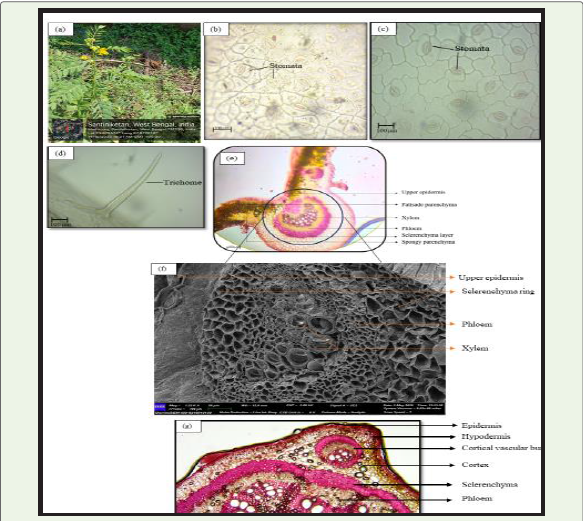
and stomata in the investigated plant species.Epidermis: Epidermal cells were observed to be irregular in shape, and the cell wall outline appeared wavy on both upper and lower surfaces. The size of epidermal cells was measured to be 35.1±1.2μm x 29.31±1.1μm on the upper surface, and 28.13±1.2μm x 12.9±1.1μm on the lower surface. The epidermal frequencies were found to be 135.21±1.22 /mm² and 156.21±1.02 /mm² on the upper and lower surfaces, respectively [Table 1] [Figure 1e].
Stomatal complex: The leaves were found to be amphistomatic,
with stomata present on both surfaces. The stomata are paracytic.
The size of stomata was measured 24.23±2.5μm x 15.30±2.5 μm
with 114.11±1.2/mm² stomatal frequency on the upper surface and
22.33±2.5 x 15.33±1.5 μm and 135.23±1.01/mm² stomatal frequency
on the lower surface. The stomatal index was 16±1.1 and 21±1.8 on
the upper and lower surfaces, respectively [Table 1] [Figure 2b,2c].
Trichomes: Unicellular, non-glandular trichomes were
observed on the upper epidermis. [Figure 2d]. The length of
trichomes was 120.12±1.61μm, and the breadth was 15.41±0.65μm.
The frequency of trichome and trichome index was measured to be
13±1.2 /mm2 and 2.31 %, respectively. [Table 3]
Vegetative anatomy:
Leaf anatomy: -The lamina of the leaf was dorsiventrally
differentiated. Both the upper and lower epidermises were uniseriate,
consisting of compactly arranged, rectangular epidermal cells with
cuticle on their outer walls. The mesophyll was distinctly differentiated
into palisade and spongy parenchyma. Two layers of cylindrical
palisade cells were present beneath the upper epidermis, followed
by 2–3 layers of loosely arranged, thin-walled spongy parenchyma
cells. In the midrib region, both epidermal layers were single-layered
and covered with a cuticle. A 3-4-layered collenchymatous tissue was
present only on the abaxial side, just below the epidermis. The ground
tissue was composed of parenchyma cells. A large, semicircular,
collateral vascular bundle is present in the middle of the midrib
[Figure 2e].Petiole anatomy: The transverse section of the petiole exhibited a plano-convex outline. The outermost layer was a uniseriate, cuticularized epidermis with compactly arranged cells. The ground tissue consisted of 2-3 layers of collenchyma cells, followed by 3-4

layers of parenchyma cells. The vascular system comprises five
vascular bundles arranged in a U-shaped arc situated within the
ground tissue [Figure 2g].
Wood anatomy: The transverse section of the wood showed vessels, Ray cells and fibres. Vessels looked like round or oval, empty pores present. Vessel distribution is mostly solitary, sometimes twin. Ray cell are parenchymatous, arranged longitudinally and rectangular in shape. Cells were thin-walled. Fibres were lignified, more or less hexagonal or polygonal in shape. They were compactly arranged along with xylem parenchyma [Figure 3A]. Transverse Longitudinal section. (T.L.S.) of wood exhibited [Figure 3B] show mostly uniseriate ray structure. The frequency of uniseriate ray
Wood anatomy: The transverse section of the wood showed vessels, Ray cells and fibres. Vessels looked like round or oval, empty pores present. Vessel distribution is mostly solitary, sometimes twin. Ray cell are parenchymatous, arranged longitudinally and rectangular in shape. Cells were thin-walled. Fibres were lignified, more or less hexagonal or polygonal in shape. They were compactly arranged along with xylem parenchyma [Figure 3A]. Transverse Longitudinal section. (T.L.S.) of wood exhibited [Figure 3B] show mostly uniseriate ray structure. The frequency of uniseriate ray

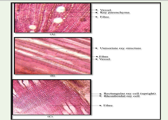
structure was 3.60±0.56/mm2. The height and width of the ray
structures were 312.01±23.23μm and 11.23±7.01 μm, respectively.
Fibres were longitudinally arranged, lignified, and closely packed on
both sides of the ray structure. Elongated fibre cell having a reduced
cell lumen with thick cell wall. The radial longitudinal section (R.L.S.)
showed heterogenous nature of ray cells having both rhomboidal
and rectangular ray cell, arranged uprightly, thin-walled and
parenchymatous. The fibres were elongated, with lignified wall and
longitudinally arranged. [Figure 3C].
Microchemical colour reaction test:
Preliminary qualitative chemical investigations of the methanol
extracts from the leaves have shown the existence of various chemical
groups such as alkaloids, flavonoids, steroids, tannins, saponins, and
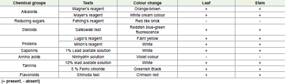
proteins, and the absence of reducing sugars and amino acids. [Table 5]Physico-chemical evaluation:
The Moisture content and ash value of the leaf and stem powder
drugs are given in tabular form [Table 6]. Moisture content for the
leaf and stem was estimated to be 10.66%. and 11.9 % respectively
whereas Ash value in leaf for total ash, acid insoluble, and watersoluble
were 6.64%, 4.67% and 1.8 %, respectively.Discussion
This study investigates the foliar micromorphology, anatomical
characteristics (leaf, petiole, and wood), preliminary phytochemical
screening and physicochemical properties, of S. sophera. The unique
features identified in this study can serve as valuable markers for
authenticating crude drugs derived from this plant and detecting
potential adulterants. Studies demonstrated the effectiveness of
foliar epidermal cell characteristics in identifying leaf based crude
drugs. In this study, the epidermal cell walls exhibited a wavy cell


wall outline, with irregularly shaped cells on both the adaxial and
abaxial surfaces. Notably, the epidermal cells on the adaxial surface
(35.1±1.2μm x 29.31±1.1μm) were larger than those on the abaxial
surface (28.13±1.2μm x 12.9±1.1μm). These distinctive epidermal cell
features provide valuable diagnostic markers for identifying the leaf
part of this plant.
Studies have shown that stomatal analysis holds significant taxonomic and pharmacognostic value in accurately identifying plant taxa, including medicinal plants (Begum and Rahaman, 2021) [1].
Our investigation revealed that S. sophera exhibits strictly paracytic stomata on both adaxial and abaxial leaf surfaces. The stomatal index, a valuable marker for taxonomic identification, was calculated to be 16±1.1% on the adaxial surface and 21±1.8% on the abaxial surface, providing a distinctive characteristic for this species. The anatomical distinction of the petiole often plays a crucial role in identifying plant taxa. The vascular system of petiole comprises five vascular bundles arranged in a U-shaped arc situated within the ground tissue, which might be a trait specific to this species.
The pharmacognostic evaluation of medicinal plants relies heavily on chemical profiling and bioactivity assessments, which provide valuable insights into their therapeutic potential (Begum and Rahaman, 2021, Anjuma et al, 2024) [1,11]. In this study, the leaf and stem extract of the investigated plant was found to contain a diverse range of phytochemicals, including alkaloids, flavonoids, steroids, saponins, and tannins. These compounds have been associated with numerous health benefits, such as antioxidant, anti-inflammatory, antimicrobial, and anticancer effects. The presence of these bioactive metabolites in the leaf extract highlights the plant’s potential as a rich source of natural products with therapeutic applications, supporting its traditional use in folk medicine and warranting further scientific investigation.
Studies have shown that stomatal analysis holds significant taxonomic and pharmacognostic value in accurately identifying plant taxa, including medicinal plants (Begum and Rahaman, 2021) [1].
Our investigation revealed that S. sophera exhibits strictly paracytic stomata on both adaxial and abaxial leaf surfaces. The stomatal index, a valuable marker for taxonomic identification, was calculated to be 16±1.1% on the adaxial surface and 21±1.8% on the abaxial surface, providing a distinctive characteristic for this species. The anatomical distinction of the petiole often plays a crucial role in identifying plant taxa. The vascular system of petiole comprises five vascular bundles arranged in a U-shaped arc situated within the ground tissue, which might be a trait specific to this species.
The pharmacognostic evaluation of medicinal plants relies heavily on chemical profiling and bioactivity assessments, which provide valuable insights into their therapeutic potential (Begum and Rahaman, 2021, Anjuma et al, 2024) [1,11]. In this study, the leaf and stem extract of the investigated plant was found to contain a diverse range of phytochemicals, including alkaloids, flavonoids, steroids, saponins, and tannins. These compounds have been associated with numerous health benefits, such as antioxidant, anti-inflammatory, antimicrobial, and anticancer effects. The presence of these bioactive metabolites in the leaf extract highlights the plant’s potential as a rich source of natural products with therapeutic applications, supporting its traditional use in folk medicine and warranting further scientific investigation.
In Pharmacognosy, physical constant of crude drug plays a very
important role in case of crude drug identification. (Sultana et al.,
2023) [3]. In this study, physical constants of the crude drugs obtained
from leaf parts of the investigated plant have been reported here
for the first time. Moisture content of a crude drug is an important
parameter in respect of its shelf life because insufficient drying favours
the growth of molds and microorganisms which ultimately spoil the
biomass and active principles of the drugs. It has also been established
that moisture content is directly related to maintain the stability and
quality of crude drugs (Wungsem, 2013, Begum and Rahaman, 2021)
[1]. The moisture content of the leaf part of the studied plant part
was found to be 10.66 %. Among the physical constants ash value
is considered as an important tool in appraisement of purity and
identity of a crude drug also. It is considered as an indicator for
mineral constituents of the crude drugs or medicinal plants. The
total amount of ash found in the leaf part (6.64 %) and in the stem
(5.59%) represent presence of inorganic minerals like carbonate,
oxalate, phosphate including silica and siliceous matters. The water soluble
ash content in leaf (1.8%) and stem (3.25%) is estimated by
measuring the amount of ash soluble in water which includes mostly
the phosphate salts and some oxalate and carbonate salts. Like total
ash, content of acid insoluble ash also provides a very confirmatory
character that helps in authentication and quality control of the
plant-based crude drug. Our result showed that the acid insoluble ash
value of the leaf part is quite low 4.67% as observed in leaf part and
2.34% in bark (Khare et al., 2017) [12-30].
Conclusion
This study provides detail anatomical and pharmacognostic
features which will helpful in identifying and authenticating the
leaf drug obtained from the S. sophera. Moreover, pharmacognostic
characters of the crude drugs will enrich the database of the studied
medicinal plants. Pharmacognostic study revealed that the leaf and
stem both parts of plant contain a good amount of therapeutically
important chemical groups such as phenolics, flavonoids, tannins,
and alkaloids. The presence of such phytochemicals in significant
amounts clearly indicates the healing potential of this medicinal plant
used traditionally in a varied range of health-related problems and
elucidates the rationale for investigating its various medicinal uses.
The overall study can lead us towards further scientific investigation
to explore the potential of this medicinal plant for the development of
bioactive natural products.
Acknowledgement
Grateful acknowledgement is extended to the Department of
Botany, Visva-Bharati (sponsored by UGC-DRS-SAP and DSTFIST),
for providing the essential laboratory facilities.
References
9. World Health Organization (1998) Quality control methods for medicinal plant materials. WHO, Geneva.
18. Metcalfe CR, Chalk L (1950) Anatomy of the dicotyledons (Vol. 1). Oxford, UK: Clarendon Press, UK.
Citation
Ghosh R, Mandal S, Roy A. Anatomical and Pharmacognostic Study of Leaf and Stem of a Medicinally Important Plant, Senna sophera (L.) Roxb. in and Around Santiniketan, West Bengal. J Plant Sci Res. 2026;13(2): 297.