A Biochemical and In Vitro Study on the Protective Effects of Quercetin on Lysosomal Glycohydrolases, Cathepsins and Adenosine Triphosphatases in Isoproterenol Induced Myocardial Infarcted Rats
Ponnian Stanely Mainzen Prince* and Balakrishnan Sathya
Department of Biochemistry and Biotechnology, Annamalai University, Annamalai Nagar-608 002, Tamil Nadu, India
Corresponding author: Dr. P. Stanely Mainzen Prince, Department of Biochemistry and Biotechnology, AnnamalaiUniversity, Annamalai Nagar- 608 002, Tamil Nadu, India, Tel No: +914144239141; Fax: +914144239141; E-mail:ps_mainzenprince@yahoo.co.in
Citation: Stanely Mainzen Prince P, Sathya B. A Biochemical and In Vitro Study on the Protective Effects of Quercetin on Lysosomal Glycohydrolases, Cathepsins and Adenosine Triphosphatases in Isoproterenol Induced Myocardial Infarcted Rats. J Enzymol Metabol. 2017;3(1): 108.
Copyright © 2017 Stanely Mainzen Prince P. This is an open access article distributed under the Creative Commons Attribution License, which permits unrestricted use, distribution, and reproduction in any medium, provided the original work is properly cited.
Journal of Enzymology and Metabolism | Volume: 3, Issue: 1
Submission: Submission: 09/04/2017; Accepted: 21/04/2017; Published: 28/04/2017
Abstract
This study was aimed to evaluate the protective effects of quercetin on lysosomal glycohydrolases, cathepsins and membrane bound adenosine triphosphatases in isoproterenol induced myocardial infarcted rats. Male albino Wistar rats were pretreated with quercetin (10 mg/kg body weight) daily for a period of 7 days. After the pretreatment period, isoproterenol (100 mg/kg body weight) was injected subcutaneously into rats twice at an interval of 24 h to induce myocardial infarction. The activities of serum creatine kinase and lactate dehydrogenase were increased significantly (P<0.05) and the activities of lysosomal enzymes (β-glucuronidase, β-N-acetyl glucosaminidase, β-galactosidase, cathepsin-B and cathepsin-D) were increased significantly (P<0.05) inthe serum and heart of isoproterenol induced myocardial infarcted rats. Isoproterenol also decreased the activities of β-glucuronidase and cathepsin-D in the lysosomal fraction and enhanced in the cytosolic fraction. The activity of sodium/potassium dependent adenosine triphosphatase was decreased significantly (P<0.05) and the activities of calcium and magnesium dependent adenosine triphosphatases were increased significantly (P<0.05) in isoproterenol induced myocardial infarcted rats. Pretreatment with quercetin (10 mg/kg body weight) showed significant (P<0.05) protective effects on cardiac marker enzymes, lysosomal glycohydrolases, cathepsins and adenosine triphosphatases in isoproterenol induced myocardial infarcted rats. The in vitro study confirmed the strong reducing property of quercetin. Thus, the results of our study reveal that quercetin minimizes isoproterenol induced myocardial damage by reinstating the activities of lysosomal enzymes and adenosine triphosphatases to near normal levels. The observed effects are due to the membrane stabilizing and reducing properties of quercetin.
Keywords: Quercetin; Isoproterenol; Myocardial infarction; Lysosomal enzymes; Membrane bound adenosine triphosphatas
Introduction
Myocardial infarction (MI) is the acute condition of necrosis of the myocardium that occurs as a result of imbalance betweencoronary blood supply and myocardial demand. Isoproterenol (ISO), a synthetic catecholamine causes severe stress in the myocardial tissue and its high doses produce MI. The induction of MI in experimental animals by ISO is probably due to its action on the sarcolemmal membrane, stimulation of adenylate cyclase, activation of sodium (Na+) and calcium (Ca2+) channels, exaggerated Ca2+ inflow and energy consumption leading to cellular death [1]. Lysosomal enzymes are crucial mediators of MI which play a vital function in the inflammatory process. In myocardial ischemia, the increased activities of glycohydrolases unremarkably diminish the lysosomal stability ensuing in necrosis of myocardium. Cathepsins are lysosomal proteases possibly involved in autophagic digestion of discrete areas of cytoplasm, myofibrillary and mitochondrial proteins. The leakage of hydrolytic enzymes from lysosomes after coronary occlusion may be a causative factor in the development of myocardial cellular injury. Adenosine triphosphatases (ATPases) of cardiac cells play a significant role in the contraction and relaxation cycles of cardiac muscle. Alterations in the properties of these ion pumps may affect cardiac function.
Dietary factors play a key role in the development of various human diseases, including cardiovascular diseases. Quercetin belongs to a group of flavonoids, naturally occurring polyphenolic compounds ubiquitously found in fruits, vegetables, herbs, tea, wine etc. It is the most commonly found flavonoid present in the human diet. Previously, we reported the protective effects of quercetin on lipids and lipoproteins in ISO induced myocardial infarcted male Wistar rats [2]. In continuation of our research on quercetin, we evaluated the protective effects of quercetin on lysosomal enzymes and membrane bound ATPases in ISO induced myocardial infarcted rats.
Materials and Methods
Experimental animals and diet
Male albino Wistar rats (Rattus norvegicus) weighing 180-200 g, obtained from the Central Animal House, Rajah Muthiah Institute of Health Sciences, Annamalai University, Tamil Nadu, India were used in this study. They were housed (3 rats/cage) in polypropylene cages (47x34x20 cm) lined with husk, renewed every 24 h under a 12:12 h light and dark cycle at around 22 °C. The rats had free access to the tap water and food. The rats were fed on a standard pellet diet (Pranav Agro Industries Ltd., Maharashtra, India). The experiment was carried out according to the guidelines of the Committee for the Purpose of Control and Supervision of Experiments on Animals, New Delhi, India and approved by the Animal Ethical Committee of Annamalai University.
Chemicals
Quercetin and isoproterenol hydrochloride were purchased from Sigma Chemical Co., St. Louis, MO, USA. p-nitrophenyl- β-D-glucuronide and α-N benzoyl-DL-arginine-p-nitroanilide hydrochloride were purchased from Himedia, Mumbai, India. All other chemicals used were of analytical grade.
Experimental protocol
The rats were randomly divided into four groups of six rats each.Group I: Normal control rats; Group II: Rats were orally treated withquercetin (10 mg/kg body weight) daily for a period of 7 days by anintragastric tube; Group III: Rats were induced MI by subcutaneous injection of ISO (100 mg/kg body weight) at an interval of 24 h for 2 days (on 8th and 9th day); Group IV: Rats were pretreated with quercetin (10 mg/kg body weight) orally by an intragastric tube daily for a period of 7 days and then injected subcutaneously with ISO (100mg/kg body weight) at an interval of 24 h for 2 days (on 8th and 9th day). Normal control and ISO control rats were received 0.5% dimethyl sulfoxide alone daily for a period of 7 days. Quercetin was dissolved in 0.5% dimethyl sulfoxide and was given to rats orally daily for a period of 7 days. The dose determination and duration of pretreatment was fixed based on our previous study [2]. At the end of the experimental period, after 12 h of second ISO injection (10th day) all the rats were anesthetized with high dose of pentobarbital sodium (60 mg/kg body weight) and then sacrificed by cervical decapitation. For serum, blood was collected in dry tubes without anticoagulant. Heart tissues were excised immediately and rinsed in ice-chilled saline.
Biochemical estimations
Separation of sub cellular fractions, assay of cardiac marker enzymes, lysosomal enzymes and membrane bound ATPases: The heart tissue samples were cut open and placed in isotonic saline to remove the blood. Then, the fresh heart tissues were homogenized in 0.25 M ice-cold sucrose solution at 4 °C. A portion of this homogenate was used to determine the total activity. Another portion of the homogenate was subjected to differential centrifugation and the different fractions were separated [3]. Myocardial sub fractions were treated with Triton X-100 (final concentration 0.2% v/v) in ice for 15 min prior to the determination of enzymic activities. Activities of serum CK and LDH were measured by standard commercial kits. The activity of β-glucuronidase in the serum, heart tissue homogenate and subcellular fractions were assayed according to the method of Kawai and Anno [4]. The activity of β-N-acetyl glucosaminidase in the serum and heart tissue homogenate was assayed by the method of Moore and Morris [5]. The activity of β-galactosidase in the serum and heart tissue homogenate was assayed by the method of Conchie et al. [6]. The activity of cathepsin-B in the serum and heart tissue homogenate was assayed by the method of Barrett [7]. Cathepsin-D activity in the serum, heart tissue homogenate and subcellular fractions were assayed by the method of Sapolsky et al. [8]. The protein content of the tissue homogenate was estimated according to the procedure of Lowry et al. [9]. The activity of Na+/K+ ATPase in the heart tissue homogenate was assayed according to the method of Bonting [10]. The activity of Mg2+ ATPase in the heart tissue homogenate was assayed according to the method of Ohnishi et al. [11]. The activity of Ca2+ ATPase in the heart tissue homogenate was assayed according to the method of Hjerten and Pan [12].
The in vitro reducing power of quercetin: The reducing power of quercetin was determined by the method of Oyaizu [13].
Statistical analysis
Statistical analysis was performed by one way Analysis of Variance(ANOVA) followed by Duncan’s multiple range test (DMRT) using Statistical Package for the Social Science (SPSS) software package version 12.00. Results were expressed as mean ± S.D. for 6 rats in each group. P values<0.05 were considered significant.
Results
Effect of quercetin on serum CK, LDH and lysosomalenzymes in the serum and heart
Rats induced with ISO showed significant (P<0.05) increased activities of serum CK and LDH (Group-III) compared to normal weight) daily for a period of 7 days significantly (P<0.05) decreased the activities of CK and LDH in the serum of ISO induced myocardial infarcted rats (Group-IV) compared to ISO alone induced myocardial infarcted rats (Group-III) (Figure 1). The activities of serum and total heart homogenate β-glucuronidase, β-N-acetyl glucosaminidase, β-galactosidase, cathepsin-B and cathepsin-D were significantly (P<0.05) increased in ISO induced myocardial infarcted rats (Group- III) compared to normal control rats (Group-I). Oral pretreatment with quercetin (10 mg/kg body weight) significantly (P<0.05) decreased the activities of these enzymes in the serum and total heart homogenate of ISO induced myocardial infarcted rats (Group-IV) compared to ISO alone induced myocardial infarcted rats (Group- III) (Figures 2a and 2b).
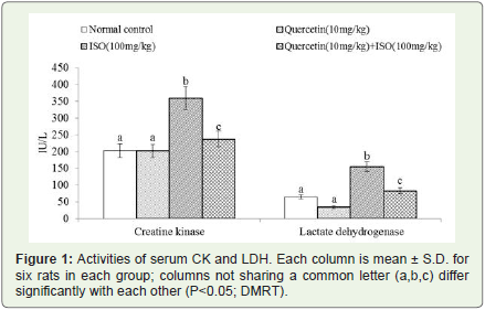
Figure 1: Activities of serum CK and LDH. Each column is mean ± S.D. for six rats in each group; columns not sharing a common letter (a,b,c) differ significantly with each other (P<0.05; DMRT).
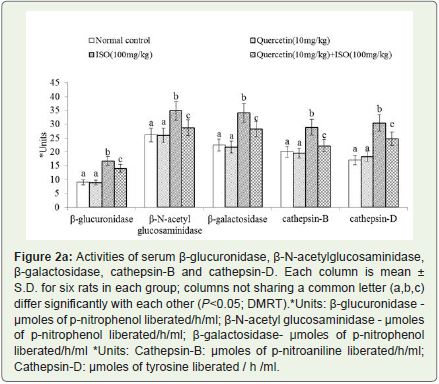
Figure 2a: Activities of serum β-glucuronidase, β-N-acetylglucosaminidase, β-galactosidase, cathepsin-B and cathepsin-D. Each column is mean ± S.D. for six rats in each group; columns not sharing a common letter (a,b,c) differ significantly with each other (P<0.05; DMRT).*Units: β-glucuronidase - μmoles of p-nitrophenol liberated/h/ml; β-N-acetyl glucosaminidase - μmoles of p-nitrophenol liberated/h/ml; β-galactosidase- μmoles of p-nitrophenolliberated/h/ml *Units: Cathepsin-B: μmoles of p-nitroaniline liberated/h/ml; Cathepsin-D: μmoles of tyrosine liberated / h /ml.
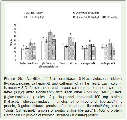
Figure 2b: Activities of β-glucuronidase, β-N-acetylglucosaminidase, β-galactosidase, cathepsin-B and cathepsin-D in the heart. Each column is mean ± S.D. for six rats in each group; columns not sharing a common letter (a,b,c) differ significantly with each other (P<0.05; DMRT).*Units: β-glucuronidase- μmoles of p-nitrophenol liberated/h/100 mg protein; β-N-acetyl glucosaminidase - μmoles of p-nitrophenol liberated/h/mg protein; β-galactosidase- μmoles of p-nitrophenol liberated/h/mg protein *Units: Cathepsin-B: μmoles of p-nitro aniline liberated/ h /100mg protein; Cathepsin-D: μmoles of tyrosine liberated / h /100mg protein.
Effect of quercetin on β-glucuronidase and cathepsin-D insubcellular fractions
The activities of β-glucuronidase and cathepsin-D was significantly (P<0.05) decreased in the lysosomal fraction and significantly (P<0.05) increased in the cytosolic fraction of the heart in ISO induced myocardial infarcted rats (Group-III) compared to normal control rats (Group-I). Pretreatment with quercetin (10 mg/kg body weight)significantly (P<0.05) increased the activity of β-glucuronidase and cathepsin-D in the lysosomal fraction and significantly (P<0.05) decreased in the cytosolic fraction of the heart in ISO induced myocardial infarcted rats (Group-IV) compared to ISO alone induced myocardial infarcted rats (Group-III) (Figures 3a and 3b).
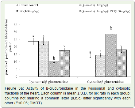
Figure 3a: Activity of β-glucuronidase in the lysosomal and cytosolic fractions of the heart. Each column is mean ± S.D. for six rats in each group; columns not sharing a common letter (a,b,c) differ significantly with each other (P<0.05; DMRT).
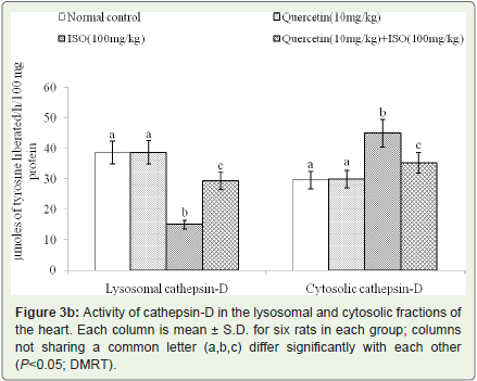
Figure 3b: Activity of cathepsin-D in the lysosomal and cytosolic fractions of the heart. Each column is mean ± S.D. for six rats in each group; columns not sharing a common letter (a,b,c) differ significantly with each other (P<0.05; DMRT).
Effect of quercetin on membrane bound enzymes in theheart
The activity of Na+/K+ ATPase was decreased significantly (P<0.05) and the activities of Ca2+ and Mg2+ATPases were increased significantly (P<0.05) in the heart of ISO induced myocardial infarcted rats (Group-III) compared to normal control rats (Group-I). Quercetin pretreatment significantly (P<0.05) increased the activity of Na+/K+ ATPase and significantly (P<0.05) decreased the activities of Ca2+ and Mg2+-ATPases in the heart of ISO induced myocardial infarcted rats (Group-IV) compared to ISO induced myocardial infarcted rats (Group-III) (Figures 4a and 4b). For all the biochemical parameters studied, administration of quercetin (10 mg/kg body weight) daily for a period of 7 days to normal rats (Group-II) did not show any significant effects compared to normal control group (Group-I).
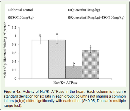
Figure 4a: Activity of Na+/K+ ATPase in the heart. Each column is mean ± standard deviation for six rats in each group; columns not sharing a common letters (a,b,c) differ significantly with each other (P<0.05; Duncan’s multiple range test).
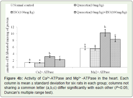
Figure 4b: Activity of Ca2+-ATPase and Mg2+ -ATPase in the heart. Each column is mean ± standard deviation for six rats in each group; columns not sharing a common letter (a,b,c) differ significantly with each other (P<0.05; Duncan’s multiple range test).
The in vitro reducing power of quercetin
Figures 5 reveals the in vitro reducing power of quercetin at different concentrations. From the results, the reducing power of quercetin increases with increasing the concentrations. The absorption at 10, 20, 30 and 40 μM concentrations of quercetin were found to be 0.15, 0.24, 0.37 and 0.49 at 700 nm respectively. Quercetin at the dose of 40 μM shows the highest absorbtion of 0.49 at 700 nm. Thus, quercetin is a potent reductant.
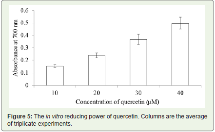
Figure 5: The in vitro reducing power of quercetin. Columns are the average of triplicate experiments.
Discussion
We observed increased activities of cardiac marker enzymes such as CK and LDH in the serum of ISO induced myocardial infarcted rats. The myocardial cells containing these enzymes are released from the heart into the circulation due to ISO induced cardiac damage. Pretreatment with quercetin (10 mg/kg body weight) near normalized the activities of these enzymes by its cardio protective activity.
Lysosomal destabilization may be prevented by restricting theleakage of lysosomal content [14]. It is possible that stabilization of myocardial cell membranes, particularly the lysosomal membranes, may prolong the viability of ischemic cardiac muscle [3]. Increased activities of lysosomal enzymes (β-glucuronidase, β-N-acetyl glucosaminidase, β-galactosidase, cathepsin-B and D) were observed in the serum and heart of ISO induced myocardial infarcted rats. Increased lipid peroxidation observed in ISO induced myocardial infarcted rats is the reason for the leakage of acid hydrolases from the enclosed sacs due to lysosomal membrane damage. Pretreatment with quercetin decreased the activities of lysosomal enzymes both in the serum and myocardium by its inhibitory effect on lipid peroxidation, thereby preventing lysosomal damage induced by ISO inducedmyocardial infarcted rats. In this context, we previously reported theantilipid peroxidation property of quercetin [2].
A compromise of lysosomal membrane integrity may lead to an undesirable elevation of enzymes in both intra and extra-cellular space, which could pave the way for cellular and tissue disorders, including apoptosis [15]. The release of β-glucuronidase is anindex of lysosomal membrane integrity. The decreased activities of β-glucuronidase and cathepsin-D in the lysosomal fraction indicate decreased stability of membranes. The elevated activities of these enzymes in the cytosolic fraction are due to the release from lysosomes to the cytosol myocardial infarcted rats. Pretreatment with quercetin enhanced the activities of these enzymes in the lysosomal fraction of ISO induced myocardial infarcted rats, thereby enhancing the stability of lysosomes by its membrane stabilizing effect.
Na+/K+ ATPase, located in the cardiac sarcolemma is considered to be involved in the maintenance of intracellular Na+ and K+ concentrations in the myocardium. Membrane Ca2+ ATPase are responsible for fine tuning of intracellular calcium as well as the contractility and excitability properties of heart muscles. Decreased activity of heart Na+/K+ ATPase is due to enhanced lipid peroxidation by free radicals on ISO induction. Enhanced heart Ca2+ ATPase activity observed in ISO induced myocardial infarcted rats is due to the activation of adenylate cyclase by ISO. Ca2+ overload in the myocardial cells during ischemia activates the Ca2+-dependent ATPase of the membrane depleting high energy phosphate stores, thereby indirectly inhibiting Na+ and K+ transport and inactivation of Na+/K+ ATPase. Further, Mg2+ ATPase activity is involved in energy requiring process in the cell and its activity is sensitive to lipid peroxidation. Pretreatment with quercetin increased the activity ofNa+/K+ ATPase and decreased the activities of Mg2+ ATPase and Ca2+ ATPase in the heart of ISO induced myocardial infarcted rats, by its membrane stabilizing property.
Further, we investigated the in vitro reducing power of quercetin.Quercetin being a phenolic compound reduces Fe3+/ ferricyanide complex to ferrous form. In our study, the reducing power of quercetin increases with increasing the concentration. Hence, quercetin is a potent reductant. The reducing power of quercetin reveals its antioxidant activity.
Conclusion
Our study revealed that pretreatment with quercetin protected the lysosomal enzymes and membrane bound ATPases in ISO induced myocardial infarcted rats. The observed effects in this study are due to the membrane stabilizing and reducing properties of quercetin. This study may be beneficial for the prevention of myocardial infarction.
References
- Devika PT, Stanely Mainzen Prince P (2008) Preventive effect of (−)epigallocatechin-gallate (EGCG) on lysosomal enzymes in heart and subcellular fractions in isoproterenol-induced myocardial infarcted Wistar rats. Chem Biol Interact 172: 245-252.
- Prince PS, Sathya B (2010) Pretreatment with quercetin ameliorates lipids, lipoproteins and marker enzymes of lipid metabolism in isoproterenol treated cardiotoxic male Wistar rats. Eur J Pharmacol 635: 142-148.
- Stanely Mainzen Prince P, Priscilla H, Devika PT (2009) Gallic acid prevents lysosomal damage in isoproterenol induced cardiotoxicity in Wistar rats. Eur J Pharmacol 615: 139-143.
- Kawai Y, Anno K (1971) Mucopolysaccharide-degrading enzymes from the liver of the squid, Ommastrephes sloani pacificus. I. Hyaluronidase. Biochim Biophys Acta 242: 428-436.
- Moore JC, Morris JE (1982) A simple automated colorimetric method for determination of N-acetyl-beta-D-glucosaminidase. Ann Clin Biochem 19: 157-159.
- Conchie J, Gelman AL, Lewy GA (1967) Inhibition of glycosidases by aldonolactones of corresponding configuration. The C-4- and C-6- specificity of beta-glucosidase and beta-galactosidase. Biochem J 103: 609-615.
- Barrett AJ (1972) A new assay for cathepsin B1 and other thiol proteinases. Anal Biochem 47: 280-293.
- Sapolsky AI, Altman RD, Howell DS (1973) Cathepsin D activity in normal and osteoarthritic human cartilage. Fed Proc 32: 1489-1493.
- Lowry OH, Rosebrough NJ, Farr AL, Randall RJ (1951) Protein measurement with the Folin phenol reagent. J Biol Chem 193: 265-275.
- Bonting SL, De Pont JJ (1981) Membrane Transport, In: Neuberger A, Van Deenen, New Comprehensive Biochemistry, Volume 2,Elsevier/North-Holland Biomedical Press, Amsterdam, The Netherlands.
- Ohnishi T, Suzuki T, Suzuki Y, Ozawa K (1982) A comparative study of plasma membrane Mg2+ -ATPase activities in normal, regenerating and malignant cells. Biochim Biophys Acta 684: 67-74.
- Hjerten S, Pan H (1983) Purification and characterization of two forms of low affinity Ca2+ -ATPase from erythrocyte membranes. Biochim Biophys Acta 728: 281-288.
- Oyaizu M (1986) Studies on Products of Browning Reaction--Antioxidative Activities of Products of browning reaction prepared from glucosamine. Jap J Nutr Diet 44: 307-315.
- Karthikeyan K, Sarala Bai BR, Niranjali Devaraj S (2007) Grape seed proanthocyanidins ameliorates isoproterenol-induced myocardial injury in rats by stabilizing mitochondrial and lysosomal enzymes: An in vivo study. Life Sci 81: 1615-1621.
- George J (2008) Elevated serum beta-glucuronidase reflects hepatic lysosomal fragility following toxic liver injury in rats. Biochem Cell Biol 86: 235-243.