Research Article
Mitogen Activated Protein Kinase Kinase-4 Upregulation is a Frequent Event in HumanStomach and Colon Cancers
Umar Mushtaq1, RafiaAnjum Baba1, Arif Ali Parray1, Hina Fayaz Bhat1, Sehar Saleem1,Usma Manzoor1, Sonaullah Kuchay2, LateefWani3 and Firdous Ahmad Khanday1*
Corresponding author: Firdous A. Khanday, Department of Biotechnology, University of Kashmir, 190006, Srinagar,Jammu and Kashmir, India, Tel: +91-990-646-2206, Fax: +91-194-242-8723,; E-mail: khandayf@kfupm.edu.sa;khandayf@kashmiruniversity.ac.in
Citation: Mushtaq U, Baba R, Parray AA, Bhat HF, Saleem S, et al. Mitogen Activated Protein Kinase Kinase-4 Upregulation is a Frequent Event in Human Stomach and Colon Cancers. J Enzymol Metabol. 2014;1(1): 103.
Copyright © 2014 Firdous Ahmad Khanday et al. This is an open access article distributed under the Creative Commons Attribution License, which permits unrestricted use, distribution, and reproduction in any medium, provided the original work is properly cited.
Journal of Enzymology and Metabolism | Volume: 1, Issue: 1
Submission: 16/08/2014; Accepted: 22/10/2014; Published: 27/10/2014
Reviewed & Approved by Dr. V. Vijaya Padma, Assistant Professor, Department of Biotechnology,Bharathiar University, India
Abstract
Mitogen-activated protein kinase kinase 4 is a dual specificity protein kinase that is encoded in humans by the MAP2K4 gene. The protein belongs to the Ser/Thr protein kinase family and has two isoforms, isoform-1(44KD) and isoform-2 (41 KD). The role of MKK4in tumors is controversial, with initial evidences suggesting as tumor suppressor role but recent studies support its pro-oncogenic role in many tissues. MKK4 has been reported as pro-oncogenic in gastric cancers but it has not been studied at protein expression level.We attempted to investigate the expression analysis of MKK4 protein infifty samples of three different types of cancers from both the sexes (28 males, 22 females) which include 28 stomach cancer samples, 18 colon cancer samples, and 12 rectal cancer sample sandcompared them with their respective normals. We found MKK4 protein expression to be up-regulated in stomach and colon cancers however analysis of rectal tumors shows no expression differences. Results observed were consistent for the chosen solid tumors when compared to their respective normals. We invoke higher expression of MKK4 in cancer might have a role to play in tumorigenesis andcould possibly be used as a novel diagnostic or prognostic marker in these cancers.
Keywords: MKK4; Cancer; Signal transduction; Stomach cancers; Colon cancers
Introduction
Gastrointestinal tract cancers are very common in Kashmiri population [1] with esophageal cancers in lead followed by stomach cancers. Also cancers of lower GIT i.e., colon and rectum have high frequency in Kashmiri population [1,2]. Cancer is a very complex disease in which signal transduction pathways is deregulated. Many proteins show differential expression in gastric and other types of cancers [3-5]. Mitogen-activated protein kinase kinases (MAPKKs) have a potential to play an important role in this disease. MAPKKs are a family of tyrosine/threonine dual specificity protein kinases, upstream of the Mitogen-activated protein kinases (MAPKs).There are four main families of MAPKs, including extra- cellular signal-regulated protein kinases (ERKs), p38-MAP Kinases, c-Jun N-terminal kinases (JNKs) and ERK5, also referred as BMK1[6,7]. MAPKKs are activated by upstream MAPKKKs that respond to a number of signaling molecules finally lead to MAPK phosphorylation and activation which initiate different cellular responses as cell proliferation, differentiation, survival, death, and transformation via phosphorylation of a number of cytoplasmic and nuclear regulatory proteins [8-10].
The MKK4 (also called JNKK1, MEK4, MAP2K4 and SEK1); a dual-specificity protein kinase is a member of the MAPK family. The protein is activated by many upstream kinases and active MKK4 mutually act with MKK7 to phosphorylate and thereby activate c-Jun NH2-terminal kinase (JNK)and p38 kinases by dual phosphorylation in the Thr-Pro-Tyr and Thr-Gly-Tyr motifs within the activation looprespectively [11-14]. The activated forms of JNKs and p38 kinases eventually initiate a wide array of biological processes, such as cell proliferation, differentiation and apoptosis [14,15]. There are two isoforms of this protein. Isoform-1 contains 399 amino acids (MW44,288 Da) and Isoform-2 contains 410 amino acids (MW 45,584Da). The sequence of isoform-2 differs from sequence the isoform-1 as follows: 39-39: G → GFQINFCEKAQS [16]. Initially MKK4 was identified as tumor suppressor protein in different cancer cell lines derived from tumors of breast, colon, lungs, pancreas, prostrate, ovaryand testis [17,18]. Normal ovarian surface epitheliums express high level of MKK4 as compared to ovarian cancer omental metastases. Reduction in the level of expression of MKK4 was observed in prostate and ovarian metastatic tissues as compared to normal prostate andovarian epithelial cells, thereby signifying that MKK4 has a metastatic suppressive role [19,21]. These findings were supported by clinical finding that MKK4 null states will encounter a growth advantages as compared to MKK-4 positive cell lines in pancreatic cancer thus lackof expression of MKK4 correlated with shorter survival time sand MKK4 positive carcinomas having half the risk of death than Mkk4- negative carcinomas [22]. Mice injected with theMKK4-/- pancreatic cancer cell line PL5 had very few lung metastases compared to those injected with the MKK4 positive or MKK4+/− cells [23]. Howeverin some cases MKK4 also functions as pro-oncogenic protein. It has been identified pro-oncogenic in breast and pancreatic cancer cell lines Ectopic expression of MKK4 in MDAMB-231 cell line by adenoviral delivery in MKK4-negative cancer lines stimulated the cellproliferation and invasion, where as knock down of MKK4 expression by small interference RNA in an MKK4-positive result in suppression of tumor growth [24]. It was also discovered that skin specific MKK4 deficient mice are resistance to carcinogen induced tumorigenesis. The mechanism by which MKK4 promotes cell proliferation and the formation of tumors is by increasing EGFR expression via the c-jun N-terminal protein kinase (JNK)/c-jun signaling pathway [25]. It was found that in MKK4 leads to over expression of JNK2 downstream, while the expression of JNK1 decreases indicates that JNK1 acts as a suppressor, while JNK2 is a promoter of skin tumor [26,27]. A pro-oncogenic function of MKK4 has been further supported by the analysis of gastric cancers and laryngeal squamous cell carcinomas. Patients with MKK4 present in tumor tissues had a significantly shorter survival compared to patients with reduced MKK4 expression[28,29].
From previous studies it has been shown that MKK4 might be involved in tumor development and metastasis progression. In quest to that, a clear systematic investigation is needed to know the expression pattern of MKK4 in different types of human cancers prevalent in Kashmir Valley. In this study we have carried out theexpression analysis of MKK4 protein in human stomach, colon and rectal cancerous tissues. This is the first demonstration of MKK4 expression in above mentioned types of cancers and this information may be used to target MKK4 protein as a potential therapeutic target.
Materials and Methods
Collection of tumor specimens
Cancer tissue samples from the patients who underwent surgical operation at Department of Surgery at Shri Maharaja Hari Singh Hospital, a total of fifty samples from both the sex (28 males, 22 females) were collected. The samples collected include 28 stomachcancer samples, 18 colon cancer samples, and 12 rectal cancer samples. All study participants gave their written informed consent. The study was also approved by the Human Ethics Review Committee of the Hospital. The adjacent normals taken for the study were obtained by dissecting about 3cm away from the tumorigenic area.
Chemicals and antibodies
Chemicals used for carrying out protein extraction and western blotting were of analytical grade and were purchased from SIGMAALDRICH (USA). From Genei laboratories (India), we purchased Bradford micro-protein estimation kit from Genei laboratories (India). PVDF membrane was purchased from Whatmann (Germany). All electrophoresis reagents were obtained from SIGMAALDRICH (USA), Thermo Scientific (USA), Qualigens (India) and Spectrochem (India), ECL solutions for protein detection wereobtained from Thermo Scientific (USA). Rabbit Monoclonal MKK4 antibody was purchased from UPSTATE-MILLIPORE, anti-rabbit HRP-conjugated Secondary Antibody and Anti-Rabbit IgG-FITC antibody was purchased from SIGMA-ALDRICH (USA). Anti-Vinculin antibody was purchased from EPITOMICS.
Western blotting
The proteins were separated using sodium dodecyl sulfate polyacrylamide gel electrophoresis (SDS-PAGE) and transferred onto a polyvinylidene fluoride membrane. The membranes were blocked in 5% non-fat milk and incubated overnight with primary antibodies. The membranes were then incubated with primary antibodies (1:1000 dilution of anti-MKK4 in 5% BSA in TBS, 1:5000 dilution of anti-vinculin Ab in 5% skimmed milk in TBS) overnightat 4 °C. After serial washing with TBS containing 0.1% Tween-20, the membranes was incubated with HRP-conjugated secondary antibodies (1:7000 dilution in 5% skimmed milk in TBS) for 2 hours at room temperature. Immunoreactive bands were visualized using ECL system (Cell Biosciences). Blots were stripped and re-probed as required.
Immunofluorescence and HE Staining
Formalin-fixed paraffin-embedded specimens were sectioned in 5μm-thick slices and sections were de-paraffinized with xylene and rehydrated with graded ethanol. Antigen retrieval was carried out byplacing specimens in citrate buffer at 98 °C for 5 min. The sections were then incubated with rabbit anti-MKK4 antibody (1:100) diluted in blocking buffer overnight, followed by incubation with secondary antibody conjugated with FITC. All the steps were followed as donepreviously [3] and the photographs of the sections were taken by a camera mounted on a Leica fluorescence microscope. Hematoxylin and Eosin staining was used as the counterstain and done as carried out previously [3].
Results
Hematoxylin and eosin staining of tissues
The tissue morphology was studied using Haematoxylin and Eosin stains (Figure 1). HE staining of tissues as expected is supposed to look different in tumor versus normal. This is so because the morphology of cancer cells is different when compared to normal cells. The structured organization as we see in the normal tissue of almost all organs of the body gets disorganized with the passage and/ or stage of cancers.
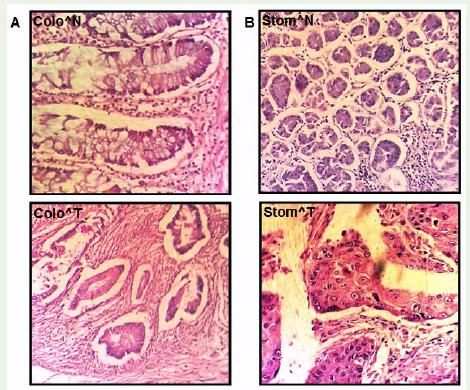
Figure 1: Hematoxylin and Eosin staining of the normal and cancerous tissues of stomach and colon.
MKK4 is up-regulated in Stomach and down-regulated in Colon cancers
In this study, we investigated the expression analysis of the MKK4 protein in stomach, colon and rectal cancers. We carried out western blot analysis, to check the expression of MKK4 protein in histologically confirmed cancerous tissues of stomach (Figure 1), colon (Figure 1) and rectal (Figure 1) cancers isolated from independent patients. Results indicate a consistent increase in MKK4 protein levels in stomach and colon cancers, when compared to their adjacent normal controls. Rectal cancers did not show any difference in the expression levels of MKK4 protein. In all the experiments, blots were stripped and treated with anti-Vinculin antibody used as loading control which showed equal expression (lower panels in allfigures) (Figure 2).
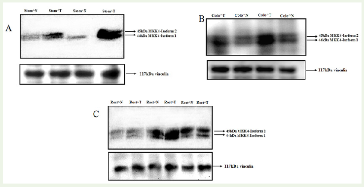
Figure 2: Up-regulation in the expression of MKK4 protein A: Representative Western blot showing the increase in the expression of MKK4in Stomach tissues(Cancerous Vs. Normal) B: Representative Western blot showing the increase in the expression of MKK4in colon tissues (Cancerous Vs. Normal) C: Representative Western blot showing the increase in the expression of MKK4in rectal tissues (Cancerous Vs. Normal).
Immunofluorescence studies reveal a higher level of MKK4 presence in Stomach and lower level of protein in Colon cancers
To further validate the results, immunostaining was also carried out alternatively in human tissues and the expression of MKK4 was found to be significantly higher in terms of recorded fluorescence signal as compared to the surrounding normal in stomach and colon cancers (Figure 3).
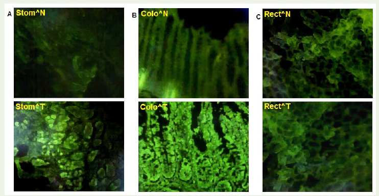
Figure 3: Immunofluorescence staining showing up-regulated expression of MKK4 protein in stomach and colon tissues (Cancerous Vs. Normal).
Discussion
The role of MKK4 in cancers is very much paradoxical and intriguing. It has been shown that skin specific MKK4 deficient mice are resistance to carcinogen induced Tumorigenesis. A pro-oncogenic function of MKK4 has been further supported by the analysis of gastric cancers and laryngeal squamous cell carcinomas. Patients withMKK4 present in tumor tissues had a significantly shorter survival compared to patients with reduced MKK4 expression. It has also been identified as proto-oncogenic in breast and pancreatic cancer cell lines[29]. Our results are in consonance with the earlier studies showing a proto-oncogenic role for MKK4 protein in cells. In the present study, we carried out Immunoblotting and Immunofluorescence of human stomach, colon and rectal tissue samples to detect the proteinexpression of MKK4.We have shown that clinical specimens of tumor tissues had increased protein expression of MKK4 in stomach and colon adenocarcinomas, but not in adjacent normal tissues, whereas the expression analysis of MKK4 in rectal samples don’t show any difference. To our knowledge, this is the first study to detect proteinexpression of MKK4 protein in stomach, colon and rectal carcinomas. We also report that MKK4 expression might be a useful prognostic marker for these type carcinomas patient survivals. Western blot analysis and Immunofluorescence techniques showed an increased MKK4 protein expression in stomach and colon cancer cases. Thereported over expression may be due to an increased transcription of the related genes or due to post-transcriptional modifications.
Acknowledgement
This work was supported by the Department of Science and Technology, Govt. of India, No: SR/SO/BB-09/2009.
Ethical clearance
The study was approved by the Human Ethics Review Committee of SMHS Hospital after obtaining patient consent and by ethical committee of University of Kashmir.
References
- Makhdoomi R, Khan AR, Besina S, Ali S, Shah BA, et al. (2011) Cancer profile in Kashmir valley an institutional experience. JK Practitioner 16: 50-54.
- Khuroo MS, Zargar SA, Mahajan R, Banday MA (1992) High incidence of oesophageal and gastric cancer in Kashmir in a population with special personal and dietary habits. Gut 33: 11-15.
- Baba RA, Bhat HF, Wani LA, Bashir M, Wani MM, et al. (2012) E3B1/ABI-1 isoforms are down-regulated in cancers of human gastrointestinal tract. Dis Markers 32: 273-279.
- Bashir M, Kirmani D, Bhat HF, Baba RA, Hamza R, et al. (2010) P66shc and its downstream Eps8 and Rac1 proteins are upregulated in esophageal cancers. Cell Commun Signal 8: 13.
- Bhat HF, Baba RA, Bashir M, Saeed S, Kirmani D, et al. (2011) Alpha-1-syntrophin protein is differentially expressed in human cancers. Biomarkers 16: 31-36.
- Pearson G, Robinson F, Beers Gibson T, Xu BE, Karandikar M, et al. (2001) Mitogen-activated protein (MAP) kinase pathways: regulation and physiological functions. Endocr Rev 22: 153-183.
- Hayashi M, Lee JD (2004) Role of the BMK1/ERK5 signaling pathway: lessons from knockout mice. J Mol Med (Berl) 82: 800-808.
- McCubrey JA, Lahair MM, Franklin RA (2006) Reactive oxygen species-induced activation of the MAP kinase signaling pathways. Antioxidants & redox signaling 8: 1775-1789.
- Dhillon AS, Hagan S, Rath O, and Kolch W (2007) MAP kinase signalling pathways in cancer. Oncogene 26: 3279-3290.
- Torii S, Yamamoto T, Tsuchiya Y, and Nishida E (2006) ERK MAP kinase in G cell cycle progression and cancer. Cancer Sci 97: 697-702.
- Sanchez I, Hughes RT, Mayer BJ, Yee K, Woodgett JR, et al. (1994) Role of SAPK/ERK kinase-1 in the stress-activated pathway regulating transcription factor c-Jun. Nature 372: 794-798.
- Tournier C, Dong C, Turner TK, Jones SN, Flavell RA, et al. (2001) MKK7 is an essential component of the JNK signal transduction pathway activated by proinflammatory cytokines. Genes Dev 15: 1419-1426.
- Boulton TG, Nye SH, Robbins DJ, Ip NY, Radziejewska E, et al. (1991) ERKs: a family of protein-serine/threonine kinases that are activated and tyrosine phosphorylated in response to insulin and NGF. Cell 65: 663-75.
- Wang X, Destrument A, Tournier C (2007) Physiological roles of MKK4 and MKK7: insights from animal models. Biochim Biophys Acta 1773: 1349-1357.
- Davis RJ (2000) Signal transduction by the JNK group of MAP kinases. Cell 103: 239-252.
- MP2K4_HUMAN UniProtKB/Swiss-Prot ( Version 145).
- Whitmarsh AJ, Davis RJ (2007) Role of mitogen-activated protein kinase kinase 4 in cancer. Oncogene 26: 3172-3184.
- Teng DH, Perry WL, Hogan JK, Baumgard M, Bell R, et al. (1997) Human mitogen-activated protein kinase kinase 4 as a candidate tumor suppressor. Cancer Res 57: 4177-4182.
- Hickson JA, Huo D, Vander Griend DJ, Lin A, Rinker-Schaeffer CW, et al. (2006) The p38 kinases MKK4 and MKK6 suppress metastatic colonization in human ovarian carcinoma. Cancer Res 66: 2264-2270.
- Yamada SD, Hickson JA, Hrobowski Y, Vander Griend DJ, Benson D, et al. (2002) Mitogen-activated protein kinase kinase 4 (MKK4) acts as a metastasis suppressor gene in human ovarian carcinoma. Cancer Res 62: 6717-6723.
- Yoshida BA, Dubauskas Z, Chekmareva MA, Christiano TR, Stadler WM, et al. (1999) Mitogen-activated protein kinase kinase 4/stress-activated protein/Erk kinase 1 (MKK4/SEK1), a prostate cancer metastasis suppressor gene encoded by human chromosome 17. Cancer Res 59: 5483-5487.
- Xin W, Yun KJ, Ricci F, Zahurak M, Qiu W, et al. (2004) MAP2K4/MKK4 expression in pancreatic cancer: genetic validation of immunohistochemistry and relationship to disease course. Clin Cancer Res 10: 8516-8520.
- Cunningham SC, Gallmeier E, Hucl T, Dezentje DA, Calhoun ES, et al. (2006) Targeted deletion of MKK4 in cancer cells: a detrimental phenotype manifests as decreased experimental metastasis and suggests a counterweight to the evolution of tumor-suppressor loss. Cancer Res 66: 5560-5564.
- Wang L, Pan Y, Dai JL (2004) Evidence of MKK4 pro-oncogenic activity in breast and pancreatic tumors. Oncogene 23: 5978-5985.
- Finegan KG, Tournier C (2010) The mitogen-activated protein kinase kinase 4 has a pro-oncogenic role in skin cancer. Cancer Res 70: 5797-5806.
- Chen N, Nomura M, She QB, Ma WY, Bode AM, et al. (2001) Suppression of skin tumorigenesis in c-Jun NH(2)-terminal kinase-2-deficient mice. Cancer Res 61: 3908-3912.
- She QB, Chen N, Bode AM, Flavell RA, Dong Z (2002) Deficiency of c-Jun-NH(2)-terminal kinase-1 in mice enhances skin tumor development by 12-O-tetradecanoylphorbol-13-acetate. Cancer Res 62: 1343-1348.
- Huang C, Huang K, Wang C, Jiang ZD, Li XX, et al. (2009) Overexpression of mitogen-activated protein kinase kinase 4 and nuclear factor-kappaB in laryngeal squamous cell carcinoma: a potential indicator for poor prognosis. Oncol Rep 22: 89-95.
- Wu CW, Li AF, Chi CW, Huang CL, Shen KH, et al. (2000) Human gastric cancer kinase profile and prognostic significance of MKK4 kinase. Am J Pathol 156: 2007-2015.