


Research Article
In Vivo Assessment of Clastogenicity of Chewable Tobacco
Ashoka CH1, Annapoorna CH2, Bhushanam M1*, Madhusudan S3 and Dakshayini PN4
1Department of Zoology, Dr. P Dayananda Pai- P. Satisha Pai GFGC, Mangalore, Karnataka. India.
2Department of Microbiology, Yenepoya Institute of Arts, Science, Commerce and Management, Mangalore, Karnataka, India.
3Professor in Biotechnology, MSCW, Maharani Cluster University, Bangalore, Karnataka. India.
4Professor in Zoology, GFGCW, Chintamani, Karnataka, India.
2Department of Microbiology, Yenepoya Institute of Arts, Science, Commerce and Management, Mangalore, Karnataka, India.
3Professor in Biotechnology, MSCW, Maharani Cluster University, Bangalore, Karnataka. India.
4Professor in Zoology, GFGCW, Chintamani, Karnataka, India.
*Corresponding author:Bhushanam M, Department of Zoology, Dr. P Dayananda Pai- P. Satisha Pai GFGC, Mangalore, Karnataka. India. E-mail Id: bhushanam.honey@gmail.com
Copyright: © 2026 Ashoka CH, et al. This is an open access article distributed under the Creative Commons Attribution License, which permits unrestricted use, distribution, and reproduction in any medium, provided the original work is properly cited.
Article Information:Submission: 10/09/2026; Accepted: 09/10/2026; Published: 13/10/2026
Abstract
The tobacco plant Nicotiana, has probably been responsible for more deaths than any other herb as it is market driven commodity of economic benefit. While the majority will likely be killed by use of cigarettes, tobacco use in other forms also contributes to worldwide morbidity and mortality. Chewing leaf tobacco is less used now as the ban is imposed on it. We tested the genotoxic potential of chewing leaf tobacco using in vivo cytogenetic test – peripheral
blood micronucleus test. Three doses of tobacco viz., 3%, 5 %, and 10 % were given for 14 days. Cyclophosphamide, an indirect acting clastogen was used as positive control agent and it was injected intraperitoneally to the animals only once. Double distilled water was used as negative control. The frequency of MNNCE was increased in tobacco treated mice with the maximum MN being induced in NCEs at 10% dose. The parameter selected to confirm the
clastogenicity of chewing leaf tobacco proved that chewing leaf tobacco at certain concentration is clastogenic.
Keywords:Chewing leaf tobacco; Peripheral blood micronucleus; Swiss albino mice; Howell–Jolly bodies.
Introduction
The tobacco plant has conquered the world as a powerful drug
in the form of cigarettes, cigars and pipes [1,2]. Nicotiana varieties
originate mainly from South America. The tobacco plant, Nicotiana,
has probably been responsible for more deaths than any other herb.
At present, tobacco smoking is causing over 3 million deaths a year
worldwide, and if current smoking trends continue the annual
mortality will exceed 10 million by around 2030. Undoubtedly,
tobacco is the most important avoidable cause of premature death
and disease in the world [3].
Smokeless tobacco is consumed without burning the
product and can be used orally and through nasal route. Oral
smokeless tobacco products are placed in the mouth, cheek or
lip and sucked (dipped) or chewed. In India, smokeless tobacco is
famous in the form of Gudakhu, Mishri, snuff, chewing tobacco,
Masher, Mawa, Sadagura. May be tobacco is the only one plant
now known world over for the overall health burden it causes
on public health. And probably this is the only one plant whose
references are made in almost every life science journals at least
once. The use of smokeless tobacco among women is increasing in
India [4]. Tobacco use is projected to kill one billion people during
the 21st century. While the majority will likely be killed by use of
cigarettes, tobacco use in other forms also contributes to worldwide
morbidity and mortality [5].
There is evidence that some tobacco and nicotine products may
pose less health hazard than cigarette smoking and so could reduce
morbidity and mortality due to smoking [6]. There is general
agreement in the scientific community that the health hazards from
low nitrosamine smokeless tobacco are lower than those of cigarette
smoking on the individual level. With respect to population impact,
there is some concern about attracting new users with reduced risk
products into the overall pool of tobacco users, whose acquired
disease risk would then offset the reduced disease burden among
smokers [7].
Micronuclei (MN), also known as Howell–Jolly bodies, were
first identified at the end of nineteenth century in red cell precursors
by William Howell, an American, and Justin Jolly, a Frenchman
[8]. They found small inclusions in the blood taken from cats and
rats. The small inclusions, were also observed in the erythrocytes of
peripheral blood from severe anemia patients [9]. The micronucleus
(MN) test is widely used for screening of different agents for their
genotoxic potential. It is a recommended bioassay in the test battery
for genotoxicity evaluation and has become increasingly popular
for regulatory acceptance. Micronucleus in peripheral blood is
used as an index for genotoxic potential. The mouse erythrocyte
micronucleus assay has been traditionally carried out using one or
two exposures to the test agent, followed by sampling at two or three
post exposure times to obtain a sample near the time of the transient
peak of micronucleated polychromatic erythrocytes (PCEs) [10].
Thus, we employed peripheral micronucleus test for investigation of
clastogenic effect of chewing leaf tobacco in Swiss albino mice.
Material and Methods
In the present investigation, Swiss albino mice (Mus musculus),
bred and maintained in the institutional animal house were used.
The animal studies were conducted after obtaining the approval from
the Institutional Animal Ethical Committee (IAEC) of Mangalore
University. Care of the animals and experimental procedures were
conducted as per the guidelines of CPCSEA, (Committee for the
Purpose of Control and Supervision of Experimentation on Animals)
India. Animals were housed in polypropylene shoe box cages bedded
with clean, dry paddy husk and kept in air-conditioned room at a
temperature of 22± 2◦C and relative humidity 50± 15 %. They were
fed with a standard pelleted diet and water ad libitum. 8–10 weeks
old animals of both the sexes with average body weight, 25 ± 2 g were
used for the experiments. In each experimental and control groups,
five animals were maintained. Cyclophosphamide (CP, CAS No.
6055-19-2; Batch No.GL3045, Asta Medica AG Germany), marketed
as Endoxan by German Remedies Ltd, Ponda, India was used as the
positive control. All other chemicals were obtained from MERCK,
SRL or HI media, India.
For the present study, three doses of tobacco viz., 3%, 5 %, and
10 % were chosen. Different doses were prepared by homogenizing
in mortar and pestle using suitable amount of distilled water. These
doses were administered orally in 0.2 ml quantity for 14 consecutive
days. Cyclophosphamide, an indirect acting clastogen as designed
and reported by Shruthi and Vijayalaxmi during 2016 [11]. (CP, 50
mg/kg b w) was used as positive control agent and it was injected
intraperitoneally to the animals only once. Double distilled water was
used as negative control.
The peripheral blood MN assay was done by using the method of Schlegel and MacGregor [12]. The blood was drawn from the tail vein and thin smears were prepared on clean grease free slides. They were fixed in absolute methanol for 10 minutes. The slides were then stained with buffered 10 % Giemsa (pH-6.8) taken in vertical coplin jars. Giemsa staining technique has been shown to give the best results for micronuclei and other biomarkers. It should be mentioned here that we filtered the buffered Giemsa using Whatman no 1 filter paper before staining the slides. A thorough washing under the running tap water was preferably carried out soon after removing from the stain to remove any stain particles adhering on the blood cells which would otherwise impede the scoring of the micronuclei. About 2000 NCE per animal were scanned for the presence of MN. The number of PCE corresponding to 2000 NCE was also determined [13].
The peripheral blood MN assay was done by using the method of Schlegel and MacGregor [12]. The blood was drawn from the tail vein and thin smears were prepared on clean grease free slides. They were fixed in absolute methanol for 10 minutes. The slides were then stained with buffered 10 % Giemsa (pH-6.8) taken in vertical coplin jars. Giemsa staining technique has been shown to give the best results for micronuclei and other biomarkers. It should be mentioned here that we filtered the buffered Giemsa using Whatman no 1 filter paper before staining the slides. A thorough washing under the running tap water was preferably carried out soon after removing from the stain to remove any stain particles adhering on the blood cells which would otherwise impede the scoring of the micronuclei. About 2000 NCE per animal were scanned for the presence of MN. The number of PCE corresponding to 2000 NCE was also determined [13].
Statistical Analysis:
The results of the experiments were expressed as the Mean % ±
SEM. The significance of differences among the groups was assessed
using one-way analysis of variance (ANOVA) test. The mean
difference between two groups were analyzed by Students t-test. For
all analyses p < 0.05 was set as the critical level of significance.Results and Discussion
Measuring numbers of micronuclei in mammalian red blood
cells is relatively straightforward because normoblasts (erythrocyte
precursor cells that reside in the bone marrow) expel their nuclei during
red blood cell development, leaving behind any micronuclei that have
formed. It therefore makes an ideal test for the detection of genome
instability in vivo that is minimally invasive and does not require the
death of the animal, meaning that it can easily be incorporated into a
phenotyping screen [14]. Polychromatic erythrocytes in mouse bone
marrow are abundant, easily recognizable, and have a lifetime of ~2
days before maturing into normochromatic erythrocytes [15].
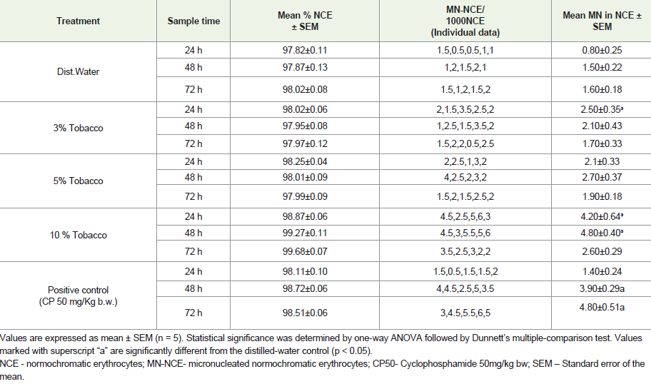
The frequency of MN NCE [Table 1]. [Figure 2] was increased in
tobacco treated mice with the maximum MN being induced in NCEs
at 10% dose. The MN-NCE in the positive control was significant only
at 48h and 72h. This may be because the positive control was treated
for only once through intraperitoneal route. The highest dose, 10%
tobacco showed MN-NCE at statistically significant levels from the
distilled water control group [Table 1].
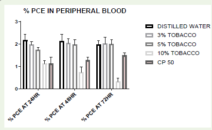
A dose-dependent decrease in the percentage frequency of PCE was found at all sampling times [Figure 1,3]. The polychromatic erythrocytes are not comparable to that found in the bone marrow. In the bone marrow, at early developmental stages the ratio of PCE to NCE is more than one. As it matures, ratio decreases slowly. The ratio of PCE to NCE decreases when an animal gets exposed to toxic agent or genotoxic compounds either through environmental contamination or through the emergency
A dose-dependent decrease in the percentage frequency of PCE was found at all sampling times [Figure 1,3]. The polychromatic erythrocytes are not comparable to that found in the bone marrow. In the bone marrow, at early developmental stages the ratio of PCE to NCE is more than one. As it matures, ratio decreases slowly. The ratio of PCE to NCE decreases when an animal gets exposed to toxic agent or genotoxic compounds either through environmental contamination or through the emergency
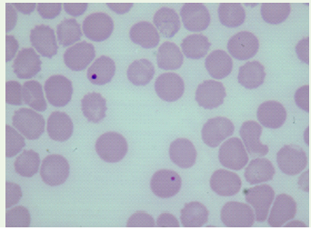
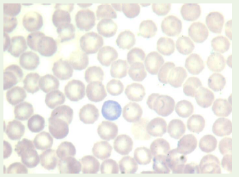
medications. In our study this effect was more pronounced in 10%
tobacco treated group. This indicates the suppressive effect of higher
dose of chewing leaf tobacco on bone marrow proliferation.
Background Micronuclei (MN) in mammalian cells serve as a reliable biomarker of genomic instability and genotoxic exposure [16]. Elevation of MN is commonly observed in cells bearing intrinsic genomic instability and in normal cells exposed to genotoxic agents. Tobacco can cause and increase the rate of nuclear anomalies in both smoking and smokeless forms compared to healthy controls. Earlier works demonstrated significant increase in the number of nucleated polychromatic erythrocytes (MN-PCE) and decreased in number of polychromatic erythrocytes (PCE) in bone marrow of nicotine-treated animals using micronucleus assay [17]. Others have investigated the in vivo micronucleus assay as part of a rat cigarette inhalation study and have also demonstrated that the level of micronuclei is not increased following cigarette smoke (CS) exposure in peripheral blood or bone marrow samples [18]. The micronuclei induction was not significantly increased following 6 weeks of cigarette smoke exposure [19].
Background Micronuclei (MN) in mammalian cells serve as a reliable biomarker of genomic instability and genotoxic exposure [16]. Elevation of MN is commonly observed in cells bearing intrinsic genomic instability and in normal cells exposed to genotoxic agents. Tobacco can cause and increase the rate of nuclear anomalies in both smoking and smokeless forms compared to healthy controls. Earlier works demonstrated significant increase in the number of nucleated polychromatic erythrocytes (MN-PCE) and decreased in number of polychromatic erythrocytes (PCE) in bone marrow of nicotine-treated animals using micronucleus assay [17]. Others have investigated the in vivo micronucleus assay as part of a rat cigarette inhalation study and have also demonstrated that the level of micronuclei is not increased following cigarette smoke (CS) exposure in peripheral blood or bone marrow samples [18]. The micronuclei induction was not significantly increased following 6 weeks of cigarette smoke exposure [19].
Erythrocyte MN represents the consequence of chromosomal
aberrations induced during the preceding mitotic divisions in
the erythroblasts [20]. Swiss albino mice are suitable for in-vivo
micronucleus assay using peripheral blood because the spleen does not
remove the micronucleate erythrocytes from the circulation, whereas
rats are not suitable. The frequency of micronucleate erythrocytes
in the peripheral blood of splenectomized rats can be used as an
index of both acute and cumulative chromosomal damage, while in
normal rats the use of peripheral blood for cytogenetic monitoring is
restricted by the selective removal of these micronucleate cells
[21-24].
Conclusions
The peripheral blood micronucleus (MN) test shows a
clear clastogenic effect in Swiss albino mice orally administered
chewing leaf tobacco for 14 days. The frequency of micronucleate
normochromatic erythrocytes (MN-NCE) was significantly higher in
the chewing leaf tobacco exposure group (3% and 5% concentrations)
than in the distilled water control group. This suggests that the
tobacco leaf’s constituents might have caused structural or numerical
chromosomal abnormalities in the mice’s bone marrow cells.
Although the relative proportion of normochromatic erythrocytes
remained steady between 97% and 98% in all treatments, the marked
increment in micronucleus induction confirms the severe clastogenic
hazard associated with sub-chronic chewing tobacco administration.
Future research is required in unraveling the therapeutic potency
of genetically modified chewing leaf tobacco in the field of
chemotherapy. This chewing leaf tobacco, though a culprit in the
society, clinically may be useful, if the pharmacological properties are
investigated and incorporated. These bioengineered Tobacco plants
may grow pharmaceuticals of the future.
Acknowledgement
The authors are very much thankful to Late Dr. K.K. Vijayalaxmi
for her former incessant support and blessings given in the true spirit
of professional recognition through the work.
References
Citation
Ashoka CH, Annapoorn A, Bhushanam M, Madhusudan S, Dakshayini PN. In Vivo Assessment of Clastogenicity of Chewable Tobacco. J Cell Sci Molecul Biol. 2026;7(1): 121.