Review Article
Detection of Leptospira in Canine Samples using Polymerase Chain Reaction based on LipL32gene and Biochemical Profiles in CanineLeptospirosis
Umesha S1*, Chandan S1, Gopinath SM2 and Shareef MI2*
Corresponding author: Umesha S, Department of Studies in Biotechnology, University of Mysore, Manasagangotri, Mysore- 570 006, Karnataka, India, Tel: +91- 0821-2419884; E-mail: umeshgroup@yahoo.co.in; su@appbot.uni-mysore.ac.in
Citation: Umesha S, Chandan S, Gopinath SM, Shareef MI. Detection of Leptospira in Canine Samples using Polymerase ChainReaction based on LipL32gene and Biochemical Profiles in Canine Leptospirosis. J Cell Sci Molecul Biol. 2014;1(1): 105.
Copyright © 2014 Umesha S et al. This is an open access article distributed under the Creative Commons Attribution License, which permits unrestricted use, distribution, and reproduction in any medium, provided the original work is properly cited.
Journal of Plant Science & Research | Volume: 1, Issue: 1
Submission: 27/03/2014; Accepted: 07/05/2014; Published: 14/05/2014.
Abstract
Canines face the risk of getting infected with commonly communicable diseases like leptospirosis, brucellosis, rabies, anthrax etc., Leptospirosis, a zoonoanthroponosis of worldwide distribution, is an acute febrile illness. Leptospirosis can be immune suppressive, predominantly human infection,commonly transmitted to human through contact of animal urine. We report here a case study of canines infected with leptospirosis, Leptospira has been isolated from blood. Leptospirosis is a septicemia bacterial disease affecting animals and human being, the infection may be symptomatic, mild or severe and acute or chronic. The clinical signals are often related to kidney disease, liver disease and other reproductive dysfunction. The disease currently diagnosed by Microscopic agglutination test (MAT). But, MAT is unable to differentiate the acute from chronic infection for the proper treatment. Since the disease affecting the kidney and the liver it is expected to have alterations in the biochemical profiles. AST and ALT in canine serum samples suspected for jaundice due to leptospirosis. In this study elevated AST and ALT were observed in positive patients. Hence, estimation of biochemical profiles may be used as an adjunct to MAT in diagnosis of acute infection. The present study was aimed to the polymerase chain reaction with the biochemical profiling for the diagnosis of leptospirosis. Biochemical and molecular techniques were performed on 40 samples of serum taken from dogs. Leptospira positives identified by dark field microscopy and PCR assay targeting partial LipL32gene of Leptospira using P23/24 primers, with a product size of 532 base pair long which are Leptospira specific. The amplicon subjected to restriction enzyme digestion using AluI, HinfI and ClaI for product of P23/24 and biochemical profile was made to correlate the biochemical AST and ALT with different clinical manifestation of leptospirosis in canines. Evidence of renal dysfunction in the present study is in conformity with the other studies. A combined effort of clinical expertise like molecular techniques along with the confirmatory laboratory back up.PCR was a reliable and precise diagnosis of leptospirosis when compare to biochemical technique in our study. The research presented here will be helpfulto improve diagnosis and control of leptospirosis in other endemic region.
Introduction
Leptospirosis is an economical important disease affecting most of the domestic animals including cattle, buffalo, dog, sheep, goat and horse along with the wild animals. As the course of leptospirosis varies from mild to rapidly fatal forms, the laboratory based techniques are important for the definite diagnosis. Culturing of the organism is the most demonstrative approach, but due its laborious condition and the consumption of time, Polymerase Chain Reaction (PCR) basedmolecular technique can be potentially rapid and specific means of diagnosis of leptospirosis especially in case of outbreak [1,2]. PCR has been used by several researchers to detect the Leptospira species in a variety of clinical specimens. In the present study, efficacy of primer capable of amplifying conserved outer membrane protein (OMP) gene i.e., LipL32. LipL32 is the major leptospiral outer membrane lipoprotein expressed during infection and is the immunodominant antigen recognized during the humeral immune response to leptospirosis in human and animals. LipL32 was tested using serum and blood samples collected from dogs from a dog shelter.
The clinical signs and severity of disease are highly up-and-down in canines. The early signs are usually unfocused and may include fever, sadness, anorexia, rigidity, myalgia, shivering and weakness. These symptoms may be followed by signs of kidney disease including anuria, increased frequency of urination, vomiting, dehydration and oral ulceration. Abortions, diarrhea, gray stools, coughing, dyspnea, conjunctivitis, weight loss and jaundice may also be seen. Some dogsdie peracutely without clinical signs. Chronic kidney disease can be a sequel. Chronic infections may be asymptomatic, or associated with fever of unknown origin and conjunctivitis. Some serovars are more likely to cause certain syndromes. Fever, hemorrhage, anemia and jaundice are typically associated with the serovar icterohaemorrhagiae. Serovar grippotyphosa tends to cause severe acute kidney failure and/or chronic active hepatitis Dogs infected with serovar pomona are often asymptomatic and chronic carriers Serovar canicola often causes chronic interstitial nephritis [3].
Cells of mammalian liver will be having an enzyme called “ALT” stands for alanine transaminase, this enzyme may also be found in a few other organs with the minimum quantities and ALT rationally liver-specific. The same enzyme is also known by an older name, SGPT, which stands for serum glutamic pyruvic transaminase. Normal serum level of ALT enzyme in canines should be between 8.2-57 u/L. ALT is known as a “leakage enzyme,” meaning the cells that contain it must die in order for it to be released. Therefore, if serum ALT levels are up, some death of liver cells has occurred. However, trends may be followed that give a suggestion of advance orworsening of liver sickness [4].
Many diseases may affect ALT, as well as non-liver conditions. For example, congestive heart failure (CHF) may result in poor blood passage, causing stagnation of liver blood flow and poor liver role.
“AST” stands for aspartate transaminase, which is also an enzyme. The older name is “SGOT,” which stands for serum glutamic oxaloacetic transaminase. AST is also present in the liver and several other organs together with skeletal muscle, heart muscle and red blood cells. Normal level in dogs is 15-66 u/L [5].
Dogs are the natural reservoir for the serogroup canicola while rodents, especially rats have that role for icterohaemorrhagiae. Leptospirosis can be identified (more easily and efficiently than with traditional bacteriological methods) by means of PCR. Using blood during the acute stage or urine or liver/kidney.
PCR-based protocols offer several advantages over standard culture techniques; the risk of cross-contamination is a major drawback. In the present study, sensitive and specific confirmative diagnostic technique for Leptospira has been developed. An attempt has been made to assess the usefulness of LipL32 gene for the diagnosisof leptospirosis over biochemical tests.
Materials and Methods
Bacterial stains, samples and media
The Leptospira suspected samples were collected from the local dog shelters, Mysore, Karnataka, INDIA. The cultures were maintained at 28-30°C in liquid medium EMJH [6] medium by routine subculture at 7-10 day intervals. The semi-solid medium with agar (0.2%) was used for maintenance of stock cultures.
Genomic DNA isolation
Template genomic DNA was extracted/ isolated from pure culture of corresponding Leptospires, and samples genomic DNA also isolated using QIAamp DNA mini kit (QIAGEN, Germany) and checked for purity using 1.5% agarose gel. It was used as template in PCR assay using primer based on LipL32gene.
Test samples
A total of 40 serum samples were collected with a history of disorders from dogs at a local dog shelter, Mysore, Karnataka, India and labeled as S1 to S40. All the samples were subjected to PCR based on LipL32 primer.
Oligonucleotide
PCR was performed using a set of primers, Primer LipL32 based primers P23 and P24 had the sequence 5’ CAA GCG CCG GAC GGT TTA GT 3’ and 5’ ACG GGC TCA CA CTG GAA TAC C 3’, respectively. The primers for amplification of LipL32 gene weredesigned from the most conserved region of LipL32 gene using molecular biology software. Primers were synthesized commercially from Chromos Biotech, Bangalore, Karnataka, India. Aliquots of the primer were prepared in nuclease free water (Promega, USA) to achieve final concentration of 25 pmol/μL.
Sensitivity and specificity of PCR primers
In order to achieve the best sensitivity and specificity for the PCRusing primer P28/29 several combinations of annealing temperatures and concentration of primers from 58 °C to 68 °C and 2.5, 5, 10, 15, 20, 25, 30, 35 and 40 pmoles respectively were tested. The following conditions were chosen: PCR was performed in 25 μl reaction volume containing 10x Taq buffer with KCl (100mM Tris HCl, 50mM KCl), 1.5mM MgCl2, 2.5mM of each dNTPs, 2U of Taq polymerase, 2.5-45 pmoles of each primer and 0.25μl of DNA (40ng/μl) extract from L. icterohaemorrhagiae. PCR chemicals were procured from Fermantas. The reactions were carried out in an Eppendorf thermocycler. Initially, PCR conditions were narrowed down by gradient PCR. The reaction started with an initial polymerase activating temperature of95 °C for 5 minutes followed 35 cycles of denaturing at 95 oC for 1 minute, annealing at 63 °C for 1 minute at G ±< 5 °C, elongation step at 72 °C for 1 minutes and a final elongation at 72 °C for 5 minutes. Finally annealing temperature was standardized to 60 °C for 1 minute.
The PCR was conducted using a 25μL reaction mixture that consists of 2.5 μL of 10x PCR buffer, 1μL of 10mM dNTP mix, 1.5 μL of 25mM MgCl2, 1.0 U of Taq DNA polymerase, 1μL (25 pmol) each of forward and reverse primers and 50 ng of template DNA. Specific PCR was performed with the following conditions: Initial denaturation at 94 °C for 5 min, followed by 30 cycles if denaturation at 94 °C for 1 min, annealing at 60 °C for 45 sec, extension at 72 °C for 30 sec and a final extension at 72 °C for 6 min. The products were checked for amplification and absence of counterfeit products by electrophoresis using 1.5% agarose gel. The gel was visualized by gel documentation system with quantity one software (Biorad, USA) and photographed.
Characterization of amplicons
The PCR product of primer P23/24 was subjected to RE digestion. The enzymes AluI (AG-CT), HinfI (G-ANTC) and ClaI (AT-CGAT) were used to characterize PCR product of P23/24. The reactions were set as under and incubated at 37 °C in a water bath for 4h. Restriction enzymes and its corresponding buffer were procured from Fermentas. ClaI and its corresponding buffer were procured from Promega. A15 ml aliquot of each digested products were mixed with 3μl of 6x loading dye (Fermentas) and loaded on to the wells of 2% agarose gel in TAE buffer. It was run at 80V for about 45 min. DNA bands were visualized under UVâ€Transilluminator and documented using gel documentation system with Quantity one software (Biorad, U.S.A).
Transformation
Ligation mixture was prepared by adding 1μl of pRSET C (50 ng) vector at the rate of and 1μl of T4 DNA ligase (3 U/ml) to 5 μl of 2x buffer. 5 μl of DNA (50 ng/ml) was added to this mixture for ligation. Ligation was done at 4 °C for overnight. Ligation mixture was added to ice cold 200μL of Top 10F’ competent cells and tapped gently, it was incubated on ice for 30 min., then Heat shock was given at 42 °C for 1 min. Immediately mixture was kept over ice for 2min. 0.8ml of LB broth was then added to it and incubated at 37 °C for 90 min in an orbital shaker. It was then centrifuged at 4000 rpm for 10 min and the pellet was plated on LB agar plate containing ampicillin (50 mg/mL) and tetracycline (50 mg/mL). The plates were then incubated over night at 37°C. Well-isolated white colonies were picked andtransferred to 5 ml of LB broth containing 5ml of ampicillin (50mg/ ml) and tetracycline (50 mg/mL). The tubes were then incubated at 37 °C in the shaker for overnight. Plasmid DNA isolation and purification was done using QIAprep spin miniprep kit (Qiagen, Germany) and microcentrifuge.
Estimation of Aspartate Transaminase (AST) and Alanine Transaminase (ALT)
Estimation of the AST, 500 μl of the reagent and 50 μl of the serum were mixed and the readings were taken in automatic photometer (M/s. Prime, USA). The total AST and ALT in the canine serum samples were estimated using the kits procured from M/S Agappe diagnostics Ltd., as per the manufacturer’s instructions. Similarly for the estimation, 500 μl of reagent and 50 μl of serum were mixed and the readings were taken in amutomatic photometer (M/s. Prime, USA).
Results
Forty samples from canines with septicemia were taken irrespective of age and sex for assessing leptospirosis disease incidence. DNA extraction from the clinical specimens by QIAamp DNA isolation kit (Qiagen, Germany) yielded a substantial amount of DNA without any protein contamination. Initially, PCR using P23/24 was standardized by carrying out reactions at various annealing temperatures ranging from 58°C to 68°C on L. icterohaemorrhagiae species which showed amplification at 58°C, 58.7°C and 60.8°C and 25 pmoles concentration of primer was standardized (Figure 1). A single band of approximately 532 bp was observed at 60 °C annealing temperature.
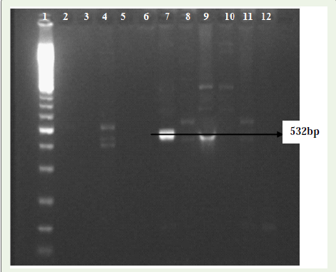
Figure 1: Electrophoresis in 1.5% agarose gel showing amplification for LipL32 gene of Leptospira species at different annealing temperature and primer concentration. Lane 1- 100bp Gene Ruler, Lanes 2-11 positive PCR product of Leptospira samples, Lane 12- Negative Control.
The primers had sufficient sensitivity to detect DNA concentration of 50-100 ng and it is specifically amplified Leptospiral DNA at 532bp (Figure 2). DNA from all the Leptospira serovars when subjected to PCR amplification, amplicons of 532 bp were observed using LipL32 specific primer. Good amplifications were evident for PCR reaction 5. carried out at the annealing temperature of 55 °C for 1 min. The 532bp amplicon was subjected to restriction digestion. The restriction enzyme pattern of this amplicon confirmed it to be the LipL32 gene of Leptospira species. All those isolates, whose identity was confirmed by digesting with restriction enzyme AluI yielded two fragments of ~240 bp and ~290 bp size, HinfI yielded two fragments of ~160 bp and ~370 bp size and ClaI yielded two fragments of ~140 bp and ~390 bp size as shown in the Figure 3
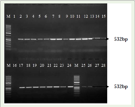
Figure 2: Electrophoresis in 1.5% agarose gel showing amplification for LipL32 gene of Leptospira species. Lane 1 and 16- Negative Control, Lanes 2-15 and 17-28 positive PCR product of Leptospira samples, M- 100bp Gene Ruler.The PCR product of the 26 positive isolates were then individually clonedinto pGMET Easy TA cloning vector and transformed into Top10 F’ E.colicompetent cells and plated on LB-ampicillin plates with IPTG and X-gal. Thediscrete colonies were then picked, cultures and plasmid was isolated forfurther use.
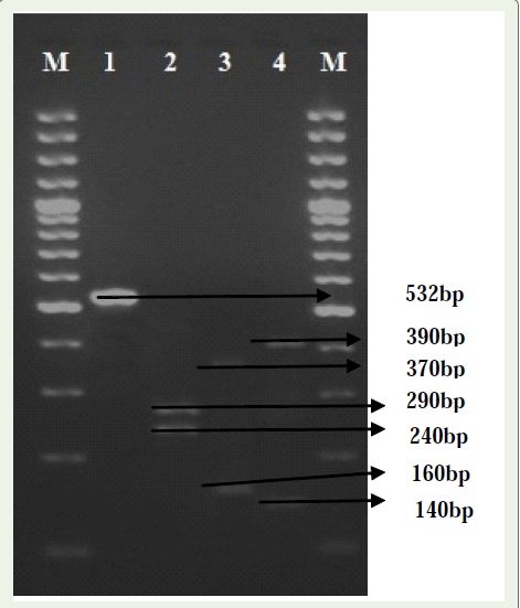
Figure 3: Agarose gel electrophoresis showing the result of restriction enzyme characterization of LipL32 gene of Leptospira. Lane M- 100 bp Gene Ruler, Lane 1- Undigested PCR product, Lane 2 to 4- PCR productsdigested with AluI, HinfI and ClaI.A total of 40 serum samples collected from suspected cases of leptospirosiswere used to estimate the antileptospiral by AST and ALT. In 40 samples,AST (>8.2 - 57 u/L) and ALT (>15-60 u/L). Samples contained elevatedlevel of AST and ALT. The increased level of ALT observed irrespective ofclinical conditions like jaundice and might be due to liver/kidney damage ormuscular damage. The determination of AST is not specific for any organ ortissue damage. However, its value is determined in liver or muscle diseasesor when a widespread damage is suspected in body. The increased level ofAST in all positive samples irrespective of clinical conditions might be due tomuscular degeneration and necrotic conditions. There is also increased levelof ALT in all the positive samples irrespective of clinical conditions might bedue to variable degree of hepatic necrosis.
Discussion
Dogs are exposed to the various rodent species in India, but the overall infection pressure also depends on the climate of each geographical area; Leptospires shed by wild animals can remain infectious for more than 6 months in temperate fresh waters (e.g. lakes, rivers and ponds). In drier areas there is less infection pressure: the incidence of canine leptospirosis in more arid regions is lower [7].
As regards the diagnosis, veterinary surgeons are generally aware of the various haematological and biochemical changes associated with acute leptospirosis. However, during the early stages of the disease, the diagnosis may be confused by a transient leucopenia (lasting 1-2 days and mimicking parvovirosis), quickly followed by the expected leucocytosis, as it is normally observed during the course of bacterial diseases. At this time a dramatic reduction in platelet count occurs, which explains the haemorrhagic lesions. During chronic leptospirosis the haematological parameters are normal, and diagnosis and assessment of the severity of the disease require serum biochemistry (for ALAT, ASAT and bile acid levels) to check for evidence of chronic hepatitis [8].
Geographical regions have not been classified by incidence, prevalence or risk of disease; this is due in part to the lack of rapid and reliable laboratory diagnostic techniques of easy use that may be practiced in non specialized laboratories around the country. Clinical symptoms of this disease do not always allow a precise diagnosis because these are highly non specific and may be mistaken with those produced by several pathogens. This makes the clinical diagnosisalone a non appropriate method that must be accompanied by laboratory tests in order to achieve accurate diagnostic results which may contribute to design of appropriate disease control strategies [9].
There was a microbiological culture negative animal that was positive in our study. A specific diagnosis of leptospirosis can be obtained by serological and molecular methods. The molecular techniques indicated the presence of pathogenic Leptospires; however these were never isolated in cultures, perhaps due to its lowconcentration in the samples. Molecular techniques can detect as low concentrate bacteria.
On the other hand, microbiological culture that is currently considered the most reliable technique for the diagnosis of Leptospira [9,10], is an expensive time consuming test that may last up to 8 weeks for results[11-13]. In this paper however, we report PCR method which is very ease and less laborious.
PCR based methods targeting different locations of Leptospiral genome for specific amplification, which hold promise as molecular tool for the diagnosis of the disease. Since conventional culture techniques are cumbersome and potentially biohazardous, they are not routinely carried out, although serological tests are preferred, but they have lesser sensitivity. Molecular methods appear to providedefinite diagnosis of this disease.
In our previous study we have shown the specificity and sensitivity of the PCR for the simultaneous detection of the potentially pathogenic Leptospira species in clinical samples. The PCR assay targeting partial sequence of LipL21 and LipL41 genes, which showed good sensitivity and specificity, a valuable tool for early diagnosis of pathogenic Leptospires directly from biological samples with clinicalsuspicion of leptospirosis.
One of the basis for using LipL32 gene based for diagnosis of leptospirosis was that this gene is conserved in all the pathogenic Leptospira species. In this paper we report the development of a PCR based test for the rapid and reliable diagnosis of Leptospira on canine blood samples, which showed 100% sensitivity and 99% specificity when compared to the biochemical culture test. These results havebeen the most accurate for the diagnosis of Leptospira in India.
Conclusion
Early diagnosis of leptospirosis is critical because of the risk ofsevere complications of the disease including pancreatitis, lung andintracranial hemorrhages, which requires intense care therapy. Theresults of the present study envisaged that PCR has advantages overother biochemical tests or microbiological tests for the early and latediagnosis of leptospiral infection and its sensitivity depends upon theclinical specimen tested. The technique holds good for screening largenumber of samples with different clinical manifestations.
Acknowledgements
The senior author (Chandan S) greatly acknowledges the financial assistance from the Indian Council of Medical Research, New Delhi, India, in the form of Senior Research Fellowship (Sanction order No. 80/673/2010-ECD-I, Dated 03.05.2011).
References
- Cheema PS, Srivastava SK, Amutha R, Singh S, Singh H, Sandey M (2007) Detection of pathogenic Leptospira in animals by PCR based on LipL21 and LipL32 genes. Indian j Exp Biol 45: 568-573.
- Chandan S, Umesha S, Bhure SK, Haraprasad N, Chandrashekar S (2011) Development of PCR assay for targeting partial LipL21 and LipL41 gene of Leptospira. Nepal Journal of Biotechnology 1: 22-30.
- Anon 2005: The center for food security and public health, College of veterinary science, Lowa State University.
- Anon 2012: AST, ALT, GGTP and Alkaline Phosphatase In Dogs’ And Cats’ Livers.
- 5.Balakrishna G, Roy GP, Thangapandian M, Govindarajan R, Ramaswamy V, et al. (2011) Biochemical profile in bovine leptospirosis. Tamilnadu J Veterinary and Animal Sciences 7: 243-246.
- Ellinghausen HC Jr, McCullough WG (1965) Nutrition of Leptopsira Pomona and growth of 13 serotype: A serum-free medium employing oleic albumin complex. Am J Vet Res 26: 39-44.
- Levette PN (2001) Leptospirosis. Clin MicrobiolRev 14: 296-326..
- Schoemaker MH, Gommans WM, Rosa LCDL, Homan M, Klok P, et al. (2003) Resistance of rat hepatocytes against bile acid-induced apoptosis in cholestatic liver injury is due to nuclear factor-kappa B activation. J Hepatol 39: 153-161.
- Agudelo -Flórez P, Retrepo M, Lotero MA (2006) Evaluation of indirect immunofluorescence assay for diagnosis of human leptospirosis. Biomedica 26: 216-223.
- Orrego A, León GDG. and RÃos B (2003) Leptospirosis risk in persons fifteen piggeries and central sacrifice of Manizales, Colombia. Archives of Veterinary Medicine 35: 205-213.
- Rahim H, Gorbanpour M, Haidari M (2005) Comparison of Leptospiral infection in the horse and donkey. Bull Vet Ins Pulaway. 49: 175-178.
- Bohorquez A, Orrego A, Giraldo Z, Mondragon M, Ramirez J, Rivera J (2002) Leptospirosis in cattle in the highland tropics of central coffee. Prevalence by direct examination and culture of urine. ACOVEZ Colombia 27: 10-16.
- Obregon M, Fernandez C, Rodriguez I, Balbis Y, Martinez B. and Rodriguez J (2004). Latex agglutination system for the rapid diagnosis of Leptospirosis Cuba. Rev Panam Salud Publica 16:259-265.