


Case Report
Delayed Cranial Progression of Pure Spinal Spontaneous Intracranial Hypotension: A Case Report
Ajit Kumar K*, Parameswaran K and Anand M
Department of Neurosurgery, Indo American Hospital Kerala, India.
*Corresponding author:Ajit Kumar K, Department of Neurosurgery, Indo American Hospital, Chemmanakary, Vaikom, Kottayam, Kerala, India. E-mail Id: dr.ajitkumar35@gmail.com
Article Information:Submission: 17/09/2026; Accepted: 05/10/2026; Published: 07/10/2026
Copyright: © 2026 Ajit Kumar K, et al. This is an open access article distributed under the Creative Commons Attribution License, which permits unrestricted use, distribution, and reproduction in any medium, provided the original work is properly cited.
Abstract
Background: Spontaneous intracranial hypotension (SIH) typically presents with orthostatic headache and characteristic intracranial MRI features. A rare pure spinal variant exists in which both symptoms and imaging abnormalities are limited to the spine. Long-term outcomes of this subtype are poorly described.
Case Presentation: A 52-year-old male initially presented with progressive upper-limb weakness without orthostatic headache. Cranial MRI was normal, while spine MRI showed ventral epidural CSF collection and venous engorgement, consistent with pure spinal SIH. He remained clinically stable for nearly five years. He later developed sudden onset headache and vomiting; imaging revealed a subacute right convexity subdural hematoma with midline shift, indicating delayed cranial progression.
Conclusion: Pure spinal SIH may appear benign initially, but delayed cranial complications can occur years later. Long-term surveillance and early treatment may prevent serious outcomes such as subdural hematoma.
Keywords:SIH; Orthostatic headache; Acute subdural hematoma
Introduction
Spontaneous intracranial hypotension (SIH) is caused by
reduced CSF pressure or volume, most commonly due to a spinal
CSF leak [1]. Typical clinical features include orthostatic headache
and characteristic intracranial MRI findings such as pachymeningeal
enhancement, venous engorgement, and brain sagging [2]. The
estimated incidence of SIH is 5 per 100,000 individuals [3].
Spinal manifestations occur in approximately 6% of cases and may present as radiculopathy or myelopathy [4], usually in conjunction with cranial symptoms. A rare clinical entity, pure spinal SIH, has been described in which the presentation and imaging abnormalities are confined entirely to the spine, with normal cranial studies [5].
Long-term follow-up data for pure spinal SIH are scarce. We present a case that remained neurologically stable for nearly five years before developing an acute subdural hematoma, representing delayed cranial progression.
Case Presentation
A 52-year-old male 5 years ago presented to neurology
department with neck pain, bilateral arm weakness and proximal
muscle atrophy. His imaging studies showed ventral epidural CSF
collection in cervical spine C3 to C5 levels. His cranial imaging at that
time was normal. A diagnosis of pure spinal SIH was established. He
was treated conservatively with bed rest, hydration, and analgesics;
no epidural blood patch was performed. His symptoms remained
stable. Now he presented with 2 days history of gradually progressive
right sided severe headache, vomiting and drowsiness. There was
no history of trauma, spinal intervention, or orthostatic headache.
Patient was drowsy (E3V4M6) and had signs of raised ICP.
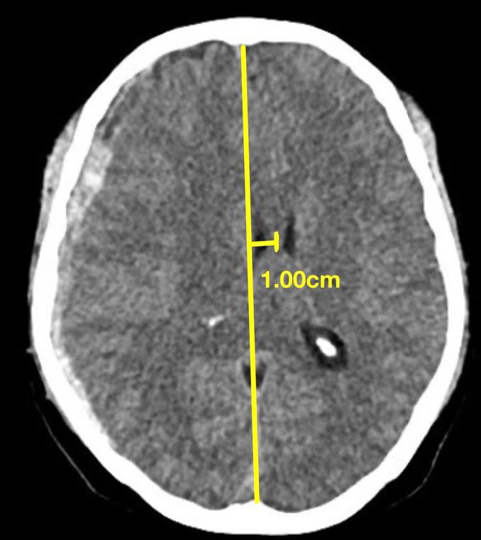
He was evaluated with CT brain which showed subacute right
cerebral convexity subdural hematoma with mass effect on right
lateral ventricle and midline shift towards left side (FIGURE-1).
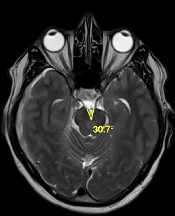
His MRI brain demonstrated reduced inter-peduncular angle of
30.7°--(normal angle measures more than 40.5°and values less
than 40.5° indicating intracranial hypotension [6]), consistent with
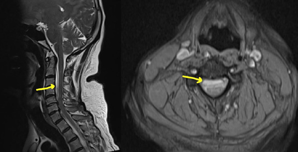
intracranial hypotension. MRI spine showed cervical spinal ventral
epidural CSF collection from C2 to C5 level.
He had Hypertension and Dyslipidaemia; other laboratory
parameters were unremarkable. The patient underwent minicraniotomy
and evacuation of subacute subdural hematoma followed
by autologous epidural blood patch. He was planned for second patch
two weeks later.
CT and MRI imaging:
Axial section of CT brain shows subacute right cerebral convexity
subdural hematoma with mass effect on right lateral ventricle and
midline shift towards left side [Figure 1]. MRI brain T2 weighted
axial section showing reduced inter-peduncular angle [Figure 2].
MRI spine T2 weighted sagittal and axial sections of cervical
spine shows ventral epidural CSF collection from C2 to C5 level
[Figure 3].Discussion
Spontaneous intracranial hypotension (SIH) is increasingly
recognized as a disorder of spinal CSF leakage rather than simply
a syndrome of low intracranial CSF pressure. Although orthostatic
headache and characteristic intracranial MRI abnormalities are the
classical manifestations, the clinical spectrum is considerably broader.
The entity described as pure spinal SIH represents an uncommon phenotype in which clinical symptoms and radiological abnormalities are initially confined to the spine, without the typical cranial manifestations of SIH[5]. Our patient demonstrated this unusual phenotype, with a cervical ventral epidural CSF collection, upperlimb weakness and muscle wasting, in the absence of orthostatic headache and with normal initial cranial imaging.
Delayed intracranial complications in pure spinal SIH are rarely reported. Chronic CSF leakage produces persistent intracranial hypotension, leading to brain descent, stretching of bridging veins, and subsequent subdural hematoma formation.
This case reinforces several key concepts:
• Normal brain MRI does not exclude future risk of intracranial
complications.
• Persistent spinal CSF leaks may lead to delayed cranial manifestations despite clinical stability.
• Early intervention-targeted epidural blood patch or surgical repair-may prevent progression to subdural hematoma.
• Long-term follow-up with periodic cranial imaging should be considered in patients diagnosed with pure spinal SIH.
Conclusion
Pure spinal SIH should not be considered benign despite the
absence of cranial findings at presentation. Significant delayed
intracranial complications such as subdural hematoma may occur
even after years of stability. Long-term monitoring and early
treatment of CSF leaks are essential to prevent morbidity.
Author Consent:
Declaration originality, competing interest, and authorship on
behalf of all authors.On behalf of all authors, I declare that this manuscript is original, has not been published previously, and is not under consideration for publication elsewhere. All authors have made substantial contributions to the work, have reviewed and approved the final version of the manuscript, and agree to its submission to the journal. The authors declare that there are no competing interests, financial or otherwise, that could have influenced the work reported in this manuscript.
References
Citation
Ajit Kumar K, Parameswaran K, Anand M. Delayed Cranial Progression of Pure Spinal Spontaneous Intracranial Hypotension: A Case Report. Indian J Neurol. 2026;7(1): 172.