Research Article
Phytochemical Composition and In vitro Anti-Hyperglycemic Potency of Eucalyptus tereticornis Bark
Asna Urooj*
Corresponding author: Dr. Asna Urooj, Professor, Department of Studies in Food Science and Nutrition, University of Mysore, Manasagangothri, Mysore, India, Telefax: +91 821 241 9632,; E-mail: atiarh@yahoo.com
Citation: Puttaswamy NY, Gunashekara DR, Ahmed F, Urooj A. Phytochemical Composition and In vitro Anti-Hyperglycemic Potency of Eucalyptus tereticornis Bark . Indian J Nutri. 2014;1(1): 102.
Copyright © 2014 Md. Asna Urooj. This is an open access article distributed under the Creative Commons Attribution License, which permits unrestricted use, distribution, and reproduction in any medium, provided the original work is properly cited.
Indian Journal of Nutrition | Volume: 1, Issue: 1
Submission: 07/07/2014; Accepted: 21/08/2014; Published: 28/08/2014
Reviewed & Approved by Dr. Sunil Misra, Scientist, Biology Division, CSIR- Indian Institute of Chemical Technology, India
Abstract
Eucalyptus tereticornis bark was screened for its anti-hyperglycemic potential by using In vitro assays viz. α-amylase, α-glycosidase and glucose adsorption. Dried powder of bark was used in glucose adsorption and alpha amylase assay, where as the water extract of the bark was used to study the inhibition of enteric enzymes (α-amylase, sucrose and α-glycosidase). The bark powder showed dose dependent amylase inhibition upto 3% sampleconcentration and further increase in concentration did not result in further inhibition, where as the water extract of the bark showed potent inhibition withIC50 value of 67.8 μg/mL. There was no notable activity observed in case of alpha-glycosidase assay. The enzyme sucrase was inhibited (35%) at 200 μg/ml extract concentration, with no further increases at higher concentrations. The bark powder showed glucose adsorption value of 1.32 mM at 10 mM glucose concentration and the adsorption increased with increase in the sample and glucose concentration. Characterization by HPLC indicated the presence of bergenin, caffiec acid, cinnamic acid and kaempferol in the aqueous extract of bark. Overall, assays partly indicate the anti-hyperglycemic potency of the bark powder which may be mediated through the inhibition of enteric enzymes. Further investigations are needed to validate this observation.
Keywords: α-amylase; Sucrase; α-glycosidase; Glucose adsorption; HPLC; Bergenin; Caffiec acid; Cinnamic acid and kaempferol.
Introduction
Diabetes is a complex metabolic disorder characterized by prolonged hyperglycemia which results due to insufficient or inefficient insulin secretion or both [1] that leads to distrubed metabolism of charbohydrates, proteins and lipds [2]. Hyperglycemia can be relieved either by increasing glucose uptake in body cells or by prolonging glucose absorption in the intestine. Latter forms the promising target to control the hyperglycemia in type-2 diabetes. Absorption of glucose in the intestine can be decreased either by controlling carbohydrate digesting enzymes or by avoiding glucose diffusion to epithelial cells or by inhibiting glucose uptake by the intestinal epithelial cells. Many synthetic drugs which are inhibitors of the carbohydrate digesting enzymes are now promoted as antihyperglycemic and thus as anti-diabetic drugs [3]. Although chemical drugs have potent carbohydrate digesting enzymes inhibiting power, they in the contrary have disadvantages of having side effects, high price and availability. In the context, medicinal plants which have comparatively less side effects, reliable and cheap, form the promisingnatural drugs to cure the disease or at least to control the disease.
Eucalyptus genus is well known in the ayurveda and other local medicine system as treatment and preventive medicine to many human ailments. This genus has been studied thoroughly for its pharmacological properties and wide array of active phytoconstituents. Eucalyptus tereticornis is one of the species of Eucalyptus genus,the plant is commonly called as Mysore gum’, ‘Mysore hybrid’ or ‘Eucalyptus hybrid. The plant is mainly used as fiber and timber source. The oil extracted from the plant is used for many ailments including muscle pain and anti-septic. The leaf of the plant shows antimicrobial activity In vitro [4,5] and anti-hyperglycemic actionin in-vivo models [6].The plant is used as one of the component in diabetic formulations in ayurveda [7]. In the context, the present study aims to evaluate Eucalyptus bark for its anti-hyperglycemic property. The assays used in the evaluation simulate the intestinal environment for carbohydrate digestion and glucose uptake.
Materials and Methods
Collection and processing of sample
Plant samples were collected from University of Mysore campus, Mysore and subsequently identified by Dr G.R Janardhana, Department of studies in Botany, University of Mysore, Mysore. The dark brown bark pieces were cut into smaller ones and dried in hot airoven at 45°C until completely dried. The dried bark was ground to fine powder and passed through 60 size mesh. Then the prepared sample was stored at 4°C until further use.
Preparation of water extract of bark
50 g bark powder was taken in 500 mL conical flask, 250 mL of distilled water was added and the conical flask was kept on shaker for 6 h at ambient temperature. The brown colored solution was centrifuged at 100 ×g to sediment water insoluble components and the supernatant obtained was filtered through filter paper. The solution obtained was freeze dried to get dark brown colored waterextracted powder. Then the extract was stored at 5°C for further use.
Preparation of crude enzymes from rat small intestine
In the preparation of crude enzymes, procedure of Dahlquist [8] was followed. Male rats of wistar strain weighing 140-160 g were obtained from central animal house, Department of Zoology, University of Mysore. The rats were sacrificed by cervical dislocation,the intestine was immediately excised. The excised intestine was washed with maleate buffer of pH 6 (0.1 M). The brush border was carefully scraped and homogenized in maleate buffer (5:1 W/V) in cold condition. Resulting homogenate was centrifuged at 1000 ×g for 15 min at 4°C. The supernatant was pipetted and stored at -20°C forfurther use as crude enzyme source of α-glycosidase and sucrase.lled the inclusion criteria were included for this study.
Phytochemical estimation
Total phenolics: Total phenolic content of the plant leaf was assayed by the method of Folin Ciocalteau [9]. In brief, various aliquots of aqueous methanolic extract of the bark (10 mg/mL) were mixed with 5 mL Folin-Ciocaleu reagent and 4 mL of sodium carbonate (75 g/L). The resulting solution was vortexed and incubated at 40°C for 30 min. The absorption was read at 765 nm. A calibration curve was prepared by using gallic acid as standard. All determinations were performed in triplicate. Total content of phenolic compounds in bark methanol extracts in gallic acid equivalents (GAE) was calculated by the following formula:
A = 0.980C + 9.925×10.3
Where, A is the absorbance of the sample and C is theConcentration as gallic acid equivalents (μg/ml).
Flavonoids: The flavonoid content was determined by apharmacopeia method using rutin as a reference compound [10]. In brief, one mL of aqueous methanolic extract in methanol (10 mg/ mL) was mixed with 1 mL aluminium trichloride in ethanol (20 g/L) and diluted with ethanol to 25 mL. The absorption at 415 nm was read after 40 min. Blank samples were prepared from 1 mL plant extract and 1 drop acetic acid, diluted to 25 mL. A standard graph was constructed using rurtin as the reference standard using the above method. All determinations were carried out in triplicate. The amount of flavonoids in plant extracts in rutin equivalents (RE) was calculated by the following formula.

Where, X- Flavanoids content (mg/g) plant extract in rutin equivalents
A-Absorbance of the Sample
Ao- Absorbance of the standard
m- Weight of the sample in mg
mo- Weight of rutin in the solution
Estimation of Total Alkaloids: The sample (0.5 g) was taken in 250 mL beaker and 200 mL of acetic acid (10%) in ethanol was added and incubated at room temperature for 10 h [11]. The resulting solution was filtered and the extract was concentrated on a water bath to one quarter of the original volume. To the concentrate, concentrated ammonium hydroxide was added drop wise to the extract until the precipitation was complete. Then, the solution was allowed to settle and the precipitate was collected, washed with dilute ammonium hydroxide and filtered. Thus resulting alkaloid precipitate was dried, weighed and expressed in alkaloids per g of the sample taken.
Saponins: The Saponin content was analyzed according to a gravimetric method previously described [12]. In brief, 250 mL of 20% ethanol was added to 10 g of the pulverized bark powder and stirred at 55°C using a magnetic stirrer for 12 h. The resulting solution was filtered using filter paper (Whatman No. 1) and the filterate volume was reduced to 40 mL under vacuum, to this 20 mL diethyl ether was added in a separating funnel and shaken vigorously. The pH of the aqueous layer was adjusted to 4.5 by adding NaOH, where as the ether layer was discarded. The pH adjusted aqueous part was extracted with 60 mL of n-butanol. The butanol extract was washed twice with 10 mL of 5% NaCl and evaporated to dryness to give a crude saponin extract which was weighed and expressed in g/g to the sample.
Tannins: Tannins were estimated according to spectrophotometric method described by Trease & Evans [13]. 5 g of the bark powder was extracted with 50 mL of boiling water and filtered. 0.5 mL of the filtrate was added to 0.5 mL of ferric solution (0.5M) in an alkaline medium and allowed to stand for 30 min for color development. The absorbance was read at 760 nm and the amount of tannin was extrapolated from a standard calibration curve for tannic acid.
HPLC analysis
The HPLC system (Waters) consisting of photodiode array detector (W2998), dual pump system (515 waters), temperature control module II (TC2 waters), pump control module (PC2 waters), system controller (EMOAA01712), and a reverse phase HPLC analytical waters Symmetry column (C18, 5 μm particle size, 4.6 × 250 mm). The mobile phase was 1% acetic acid in water (solvent A)and acetonitrile: methanol (solvent B, ratio 1:1) and the separation was performed by the following linear gradient: 70:30 (Solvent A:B) for 10 min, 60:40 (solvent A:B) for another 10 min and the flow rate was adjusted to 0.6 mL/min. Injection volume was 20 μL. The chromatogram of water extract was compared with the individualchromatogram of standards bergenin, caffiec acid, cinnamic acid and kaempferol (Sigma Chemical Co, St. Louis, USA). Data analysis was done using Empower software.
Sucrase assay
The sucrase assay was carried out according to the procedure described by Honda [14]. In brief, 10 μL of crude enzyme solution (prepared as described in section 2.3), 10 μL of sample (water extract of the bark, 10 mg/mL) and 100 μL of maleate buffer were mixed in a test tube and incubated at 37°C for 10 min. The reaction was started by adding 100 μL of substrate (60 mmol of sucrose) and incubated for 30 min at 37°C in a water bath. The reaction was stopped by heating the contents on a boiling water bath for 10 min. The liberated glucose was then estimated by GOD-POD method. A control sample was also run by following the exact procedure but without adding sample. The activity, as percent inhibition of sucrase was calculated using the formulae below.
Alpha amylase assay
The alpha amylase enzyme assay was carried out according to the procedure defined by Ou et al [15]. Effect of Bark powder on activity of enzyme was assayed using enzyme starch system. Bark powder (1-4% w/v) and 100 mg of alpha amylase enzyme powder were added to 25 mL of potato starch solution in 50 mL plastic tubes and mixed well using vortex. The tubes were incubated at 37°C for 60 min in a shaking water bath. After incubation 2 mL of 0.1 M NaOH was added and heated on a boiling water bath for 10 min to stop the enzymatic reaction. The tubes were cooled and liberated glucose was measured using GOD-POD method. A control was also run without adding any test sample. The amylase inhibitory activity in percentage was calculated using the below formula.
Alpha-glucosidase assay
The glycosidase assay was carried out according to the procedure described by Honda [14]. Crude enzyme (solution prepared as described in section materials and methods), sample (water extract of bark powder), and maleate buffer were mixed in a test tube andincubated at 37°C for 10 min. The enzyme reaction was started by adding 200μL of substrate (p-nitrophenyl-a-D-glucopyranoside 2 m mol) and incubated at 37°C. After 30 min, the reaction wasterminated by treating the mixture with boiling water for 5 min. After the addition of 1.0 mL of disodium hydrogenphosphate (0.1 M), the absorbance of the liberated p- nitrophenol was read at 400 nm. An untreated enzyme solution was used as the control.
Glucose adsorption assay
Glucose adsorption assay was carried out according to method of Ou et.al [15]. Bark powder at various concentrations (1, 2, 3 and 4%) were added to 25 mL of glucose solution at various concentrations (5, 10, 15, 20 and 25 m mol) in 50 mL volume centrifuge tubes. The tubes were incubated on a shaking water bath for 6 h at 37°C.After incubation, tubes were centrifuged at 2500 ×g for 5 min. The remaining glucose in the supernatant was measured using GODPOD method. A control was also run without adding any test samples. Glucose adsorbed by the sample was calculated by the following relation mentioned below.
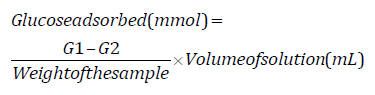
Where, G1 glucose concentration of original solution and G2 is glucose concentration of the same solution after incubation for 6h.
Statistical analysis
All the values are mean ± SD of triplicates determinations of three experiments. The data was analyzed by ANOVA followed by Tukey’s multiple comparisons test for significant differences using SPSS 14.0 software and the graphs were plotted using Origin 6.1 software. Thevalues were considered significant at p ≤ 0.05. IC50 value for all the enzyme assays were calculated by slope equation from the scatter graph in Microsoft excel.
Results
Phytochemical analysis
Values of phytochemical quantification are presented in Table 1. The bark contains significant amount of polyphenols. Flavanoid content was higher when compared to other constituents.
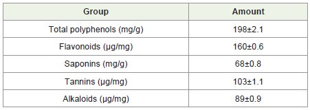
Table 1: Phytochemical composition of eucalyptus bark.
HPLC analysis
Figure 5 shows the separation pattern of phytochemicals present in aqueos extract of bark. The resolved peaks of bergenin, caffiec acid,cinnamic acid and kaemferol are identified by comparing with the chromatogram of standards. Peak area of the bergenin was higher than other resolved peaks indicating its higher percentage in the extract, followed by caffiec acid, cinnamic acid and kaempferol.
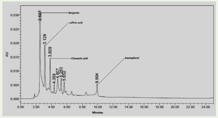
Figure 5: HPLC profile of aqueous bark extract.
Alpha amylase assay
The Figure 1 shows the values of alpha amylase inhibition by bark powder. About 33% of enzyme inhibition was noted at 1 % sample concentration and at 2% concentration a significant increase in inhibition was observed (51%), but at 3% sample concentration therewas only a slight increase in the inhibition in comparison with the former concentration, and there was no further increase in inhibition with increase in the sample concentration was observed.
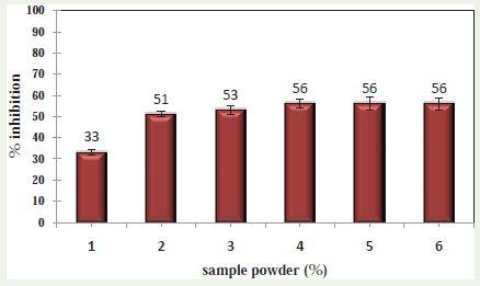
Figure 1: Effect of E. tereticornis bark powder percentage on percentinhibition of alpha amylase.
The Figure 2 shows the values of alpha amylase inhibition by water extract of bark. The extract showed dose dependent inhibition of alpha amylase, lowest activity was noted at 50 μg/mL and the activity was ceased to increase above 200 μg/mL. IC50 value of the extract as depicted by the scatter graph was found to be 64.78 μg/mL.
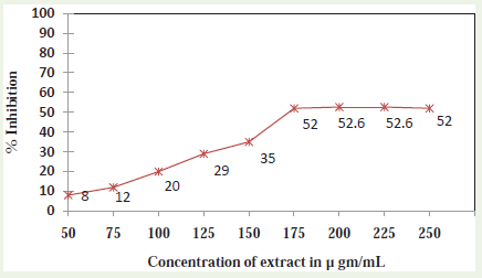
Figure 2: Effect of different concentration of E. tereticornis bark powderextract on percent inhibition of alpha amylase activity.
Alpha glycosidase
The bark water extract has no effect on the activity of alpha glycosidase.
In this observation 68% male and 32% female were admitted where 58% were rural and 42% urban living. The patients parents were illiterate 62%, primary 24%, secondary 12%, above 2%, who practices early weaning 46%, late weaning 42%, poor socio-economic population was at risk as with associated problem with tuberculosis7%, acute respiratory infection 25%, diarrhea 39%, Septicemia 29% in PEM.
Sucrase
The water extract of bark exerted significant inhibitory activity on the sucrase. The values (Figure 3) indicate that the increase in concentration of the extract results in linear increase in inhibition. There was 35% of inhibition on the activity of sucrase can be notedat 200 μg/mL.
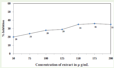
Figure 3: Sucrase enzyme inhibition activity.
Glucose adsorption
Values of glucose adsorption potency of the bark powder at various concentrations of glucose and the bark powder is shown in Figure 4. The values indicate that the glucose adsorption increase with the increase in glucose concentration as well as the sample concentration.
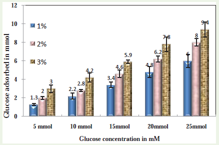
Figure 4: Glucose adsorption by Eucalyptus powder.
Discussion
Carbohydrate in the diet consists of polysaccharides and simple monosaccharide. Simple carbohydrates such as glucose, fructose,galactose are directly taken up by intestinal epithelial cells [16]. The complex polysaccharides are required to be digested into simpler sugar unit prior absorption. Digestion takes place solemnly with the help of enzymes [17]. Enzymes such as α-amylase, α-glycosidase, sucrase and lactase play a crucial role in complete digestion ofpolysaccharides into simpler monosaccharide units. The liberated mono-saccharides diffuse to intestinal epithelial cells where they are taken by the passive diffusion, facilitated diffusion through transporters named Glut transporters and by co-transport with otherions, mainly sodium ion [18]. The components which interfere with one of the above important processes can act as anti hyperglycemic agents by avoiding entry of glucose into blood stream.
Glucose adsorption is the physical phenomenon where glucose molecules get adsorbed on to the solid surface. The adsorption of glucose can result in decreased free glucose in the solution, thus can inhibit the diffusion of glucose which in turn can inhibit glucose uptake into blood [19]. The Eucalyptus tereticornis bark has good glucose adsorbing value there by indicating its capacity to adsorb glucose in the intestine and thus decreasing its availability to diffusion into blood stream. Fiber is one of the main components in the bark of the most plants [20], and the insoluble fiber has shown to havepotent glucose adsorbing potency [21], thus fiber content in the eucalyptus bark powder may be component responsible for glucose adsorption. Inhibition of α-amylase decreases the starch breakdown and thus breakdown of complex carbohydrate into simple absorbable sugars [22]. Studies report the flavonoids present in the medicinalplants strongly inhibit the alpha amylase [23], previous report on the phytochemical composition of bark of E. terticornis shows the presence of flavanoids and the present study quantitates the flavanoid content of bark. HPLC analysis indicates the presence of kaempferol, a flavonoid, in significant amount, previous study shows the significant alpha-amylase inhibitory potency of Kaempferol [24], thus the alphaamylase inhibition by the extract may be attributed by the flavanoid content, in particular by kaemperol. Very low IC50 values for extract for α-amylase inhibition indicate the potent inhibition of α-amylase by the extract. Sucrase is the enzyme involved hydrolysis of sucrose and maltose into glucose and fructose by an alpha-D-glucosidasetype action. It is one of the important enzymes involved in complete digestion of polysaccharides into simple monosacharides. Its inhibition hinders the complete digestion of complex carbohydrates into simpler monosaccharide. Inhibition of sucrose by the extract can control the disaccharide digestion into simpler monosaccharides. The water extract of the bark shows moderate sucrase inhibition, which can result in further hampering of available glucose in the intestineand thus controlling glucose entry into blood stream.
HPLC chromatogram indicates the presence of bergenin,cinnamic acid, caffiec acid along with kaempferol. Bergenin is present in higher amount in the extract. These phytochemicals have been shown to posses important role in improving blood glucose level, insulin secretion and other potential to modulate the physiologicalprocesses which are beneficial in ameliorating the diabetes pathology.
Conclusion
The results of In vitro assays are indicative of anti-hyperglycemic potency in the Eucalyptus tereticornis bark which may be attributedto the presence of important phytochemicals. The study is in the direction to study other physiological effects of the bark in respect to diabetes using In vitro and in-vivo models, and to assign safety of the bark powder using in-vivo models.
References
- Diagnosis and Classification of Diabetes Mellitus. Diabetes Care (2009) American Diabetes Association 32: 62-67.
- Bergman RN (1989) Lilly lecture 1989. Toward physiological understanding of glucose tolerance, Minimal model approach. Diabetes 38: 1512-1527.
- Tripathi BK, Srivastava AK (2006) Diabetes mellitus: Complications and therapeutics. Med Sci monit 12: 130-147.
- Badrunnisa S, Pai VR, Shantaram M (2011) Antibacterial activity of Eucalyptus tereticornis extracts for in use coolants of steel industry. International Journal of Research in Pharmaceutical and Biomedical Sciences 2: 1789-1794.
- Jain P, Nimbrana S, Kalia G (2010) Antimicrobial Activity And Phytochemical Analysis of Eucalyptus Tereticornis Bark And Leaf Methanolic Extracts. International Journal of Pharmaceutical Sciences Review and Research 4: 126-128.
- Shahraki A, Shahraki M (2013) The Comparison of Eucalyptus Aqueous Extract and Insulin on Blood Sugar and Liver Enzymes in Diabetic Male Rats. Zahedan Journal of Research in Medical Sciences 15: 25-28.
- Prabhakar PK, Doble M (2008) A target based therapeutic approach towards diabetes mellitus using medicinal plants. Curr Diabetes Rev 4: 291-308.
- Dahlqvist A (1962) Method for assay of intestinal disaccharides. Anal Biochem 7: 18-25.
- Singleton VL, Rossi JA (1965) Colorimetry of total phenolics with phosphomolybdic-phosphotungstic acid reagents. American Journal of Enology and Viticulture 16:144-158.
- State Pharmacopeia of USSR (1989) Moscow. Medicina 2: 324-334.
- Harborne JB (1984) Phytochemical methods. London; Chapman and Hall 49-188.
- Trease GE, Evans WC (1978) Pharmacology. London; Bailliere Tindall Ltd:11th Ed. 60-75.
- Obadoni BO, Ochuko PO (2002) Phytochemical studies and comparative efficacy of the crude extracts of some homeostatic plants in Edo and Delta States of Nigeria. Global Journal of Pure and Applied Sciences 8: 203-208.hers nutritional related KAP. Ind J Paed 58: 269-274.
- Honda M, Hara Y (1993) Inhibition of rat small intestinal sucrase and α-glucosidase activities by tea polyphenols. Bioscience, Biotechnology and Biochemistry 57: 123-124.
- Ou S, Kwok K, Li Y, Fu L (2001) In vitro study of possible role of dietary fiber in lowering postprandial serum glucose. J Agric and Food Chem 49: 1026-1029.
- Cirillo VP (1962) Mechanism of glucose transport across the yeast cell membrane. J Bacteriol 84: 485-491.
- William Ganong F. Review on medical physiology. California. Lange medical publications/los Altos. Section V.
- Mike Mueckler (1994) Facilitative glucose transporters. Eur J Biochem 219: 713-725.
- Gallagher AM, Flatt PR, Duffy G, Abdel-Wahab YHA (2003) The effects of traditional antidiabetic plants on in vitro glucose diffusion. Nutrition Research 23: 413-424.
- Madison. Bark and its possible uses. U.S. department of agriculture forest service forest products laboratory..
- Adiotomre J, Eastwood MA, Edwards CA, Brydon W (1990) Dietary fiber; in vitro methods that anticipate nutrition and metabolic in humans. Am J Clin Nutr 52: 128-134.
- Salt WB, Schenker S (1976) Amylase-Its Clinical Significance: A Review of the Literature. Medicine 55: 269-289.
- Tadera K, Minami Y, Takamatsu K, Matsuoka T (2006) Inhibition of alpha glucosidase and alpha amylase by flavonoids. J Nutr Sci and Vitaminol 52: 149-153. Pharmaceutical and Biomedical Sciences 2: 1789-1794.
- Kim JS, Kwon CS, Son KH (2000) Inhibition of alpha glucosidase and amylase by luteolin, a flavonoid. Biosci Biotechnol Biochem 64: 2458-2461.