Research Article
A Prospective Observational Study Comparing IOTA Simple Ultrasound Rules and RMI-2 Scoring System in Correlation with Histopathological Examination in Diagnosing Adnexal Masses at a Tertiary Care Centre
Sayantani Bhattacharyya and Falguni Patel*
Department of Obstetrics and Gynaecology, Government Medical College, Surat, Gujarat, India.
*Corresponding author:Falguni Patel, Department of Obstetrics and Gynaecology, Government Medical College, Surat, Gujarat, India. E-mail id: shivshakti12.patel@gmail.com
Article Information:Submission: 02/04/2026; Accepted: 12/05/2026; Published: 15/05/2026
Abstract
Background: Adnexal masses are frequently encountered in gynecological practice and often present a diagnostic challenge in differentiating benign from malignant lesions before surgery. Accurate preoperative assessment is essential for appropriate referral, surgical planning, and improving patient outcomes. The International Ovarian Tumor Analysis (IOTA) Simple Ultrasound Rules and Risk of Malignancy Index-2 (RMI-2) are commonly used tools for this purpose.
Objectives: To compare the diagnostic accuracy of IOTA Simple Ultrasound Rules and RMI-2 scoring system in differentiating benign and malignant adnexal masses, using histopathological examination as the gold standard. Methods: This prospective study included 50 women with adnexal masses at a tertiary care centre. Patients underwent clinical evaluation, ultrasonography, serum CA-125 testing, and assessment by IOTA Simple Rules and RMI-2, with final diagnosis confirmed by histopathology. Diagnostic performance was compared using sensitivity, specificity, PPV, and NPV.
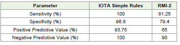
Results: IOTA Simple Rules demonstrated 100% sensitivity, 96.8% specificity, 93.75% PPV, and 100% NPV. RMI-2 showed 81.25% sensitivity, 79.4% specificity, 65% PPV, and 90% NPV. IOTA Simple Rules showed superior diagnostic performance compared to RMI-2 in differentiating benign from malignant adnexal masses.
Conclusion: IOTA Simple Ultrasound Rules are more accurate than RMI-2 in the preoperative evaluation of adnexal masses and can be used as a simple, reliable, and effective screening tool in routine gynecological practice.
Objectives: To compare the diagnostic accuracy of IOTA Simple Ultrasound Rules and RMI-2 scoring system in differentiating benign and malignant adnexal masses, using histopathological examination as the gold standard. Methods: This prospective study included 50 women with adnexal masses at a tertiary care centre. Patients underwent clinical evaluation, ultrasonography, serum CA-125 testing, and assessment by IOTA Simple Rules and RMI-2, with final diagnosis confirmed by histopathology. Diagnostic performance was compared using sensitivity, specificity, PPV, and NPV.
Results: IOTA Simple Rules demonstrated 100% sensitivity, 96.8% specificity, 93.75% PPV, and 100% NPV. RMI-2 showed 81.25% sensitivity, 79.4% specificity, 65% PPV, and 90% NPV. IOTA Simple Rules showed superior diagnostic performance compared to RMI-2 in differentiating benign from malignant adnexal masses.
Conclusion: IOTA Simple Ultrasound Rules are more accurate than RMI-2 in the preoperative evaluation of adnexal masses and can be used as a simple, reliable, and effective screening tool in routine gynecological practice.
Keywords:Adnexal mass; IOTA; RMI-2; Ovarian malignancy; Ultrasonography; Histopathology; CA-125
Introduction
Adnexal masses are common and range from benign cysts to
malignant tumors, making accurate preoperative diagnosis essential
as management varies by lesion type. These lesions may arise from the
ovaries, fallopian tubes, or surrounding structures and may present
with symptoms such as pelvic pain, abdominal distension, or may
be incidentally detected during imaging for unrelated conditions.
Accurate preoperative differentiation between benign and malignant
adnexal masses is essential for appropriate clinical management, as
it directly influences surgical planning, the extent of intervention,
and the need for referral to oncologic centers. Misclassification may
result in unnecessary extensive surgery for benign lesions or delayed
treatment in malignant cases, adversely affecting patient outcomes
Although less common, ovarian malignancies have high
morbidity and mortality due to late presentation, with vague
symptoms like abdominal pain, distension, bloating, urinary issues,
and menstrual irregularities hindering early diagnosis. Most cases are
diagnosed after the disease has spread beyond the pelvis, resulting
in poor survival outcomes, with a five-year survival rate of less than
45%. This highlights the urgent need for reliable, accessible, and costeffective
diagnostic methods for early detection and risk stratification
of adnexal masses. Given that these masses occur across all age
groups, from reproductive to postmenopausal women, an effective
and standardized approach to evaluation is critical
To improve preoperative diagnosis, several models and scoring
systems have been developed. The Risk of Malignancy Index
(RMI) is a widely used composite score based on menopausal
status, ultrasonographic findings, and serum CA-125 levels. The
IOTA Simple Ultrasound Rules, on the other hand, are based on
standardized sonographic morphological features and classify masses
as benign, malignant, or inconclusive.
The performance of these tools may vary by patient and tumor factors; thus, this study compared the diagnostic accuracy of IOTA Simple Rules and RMI-2 in adnexal masses, with correlation to histopathology.
The performance of these tools may vary by patient and tumor factors; thus, this study compared the diagnostic accuracy of IOTA Simple Rules and RMI-2 in adnexal masses, with correlation to histopathology.
Aims and Objectives
Primary Objective:
To compare the diagnostic accuracy of IOTA Simple Ultrasound
Rules and RMI-2 scoring system in differentiating benign and
malignant adnexal masses.Secondary Objective:
To correlate the findings of both scoring systems with
histopathological examination, considered the gold standard.Materials and methods
Study Design and Setting:
This was a prospective observational study conducted in
Department of Obstetrics and Gynaecology GMCS, GUJARAT
among women diagnosed with adnexal masses and planned for
surgical management.Study Population:
A total of 50 patients with adnexal masses were included in the
study.Inclusion Criteria
• Women diagnosed with adnexal masses on clinical and/or ultrasonographic evaluation
• Patients admitted for operative management
• Patients willing to participate in the study
Exclusion Criteria
• Patients managed conservatively
• Patients with incomplete investigations or unavailable histopathological diagnosis
• Patients unwilling to participate
• Ectopic pregnancy
Study Procedure
After obtaining informed consent, all eligible patients underwent:
• Detailed clinical history and physical examination
• Routine laboratory investigations
• Serum CA-125 estimation
• Ultrasonographic evaluation of adnexal mass
• Application of:
o IOTA Simple Ultrasound Rules
o RMI-2 scoring system
The final diagnosis in all cases was established by histopathological examination, which was considered the reference standard.
IOTA Simple Rules
The IOTA Simple Rules were applied using predefined sonographic benign features (B-features) and malignant features (M-features). Lesions were categorized as:
• Benign
• Malignant
• Inconclusive
RMI-2 Scoring
The Risk of Malignancy Index-2 (RMI-2) was calculated using the formula:
RMI-2 = U × M × CA-125
Where:
• U = Ultrasound score
• M = Menopausal score
• CA-125 = Serum CA-125 level (U/mL)
Outcome Measures
The diagnostic performance of both methods was assessed by:
• Sensitivity
• Specificity
• Positive Predictive Value (PPV)
• Negative Predictive Value (NPV)
Statistical Analysis
Data were entered into a Microsoft Excel spreadsheet and analyzed using appropriate descriptive and inferential statistical methods. Histopathological examination was taken as the gold standard for comparison.
Results
Demographic Characteristics:
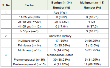
Among the 50 study participants, the majority belonged to the
26-40 years age group (58%), followed by 41–55 years (24%). Most
patients were premenopausal (70%), while 30% were postmenopausal.
Nulliparous women constituted the largest parity group.Clinical Presentation:
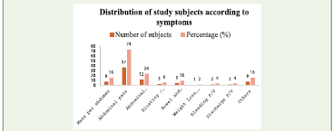
The most common presenting complaint was abdominal pain
(74%), followed by abdominal distension (24%) and mass per
abdomen (16%). Other symptoms included bloating, menstrual
disturbances, bowel/bladder complaints, and vaginal bleeding/
discharge.Histopathological Findings:
Histopathology showed most adnexal masses were benign,
with fewer malignant cases. Benign lesions included nonneoplastic
and benign neoplastic masses (e.g., serous and mucinous
adenomas), while malignant lesions included serous and mucinous
cystadenocarcinoma, endometrioid carcinoma, dysgerminoma, and
granulosa cell tumor.IOTA Classification
According to IOTA Simple Rules:
• 31 cases (62%) were classified as benign
• 16 cases (32%) were classified as malignant
• 3 cases (6%) were classified as inconclusive
Diagnostic Accuracy:
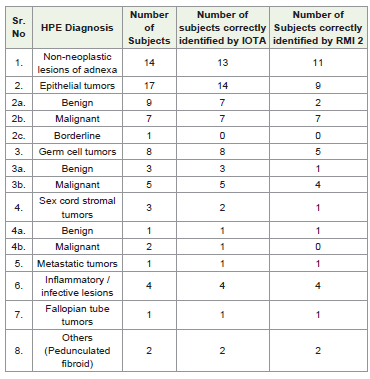
HPE distribution of study subjects and comparison of RMI2
with IOTA Simple USG Rules among the 50 study subjects. 14 cases
of non-neoplastic lesions of adnexa are identified. In the Epithelial
tumors of ovary: 9 Benign forms are presented as Serous adenoma
and Mucinous adenoma [6]; 7 Malignant cases are presented as
Serous cystadenocarcinoma [5], Mucinous cystadenocarcinoma [1]
and Endometroid cancer [1]. Among Germ cell tumors, 5 Malignant
cases are presented as Dysgerminoma (4), Mixed Germ cell tumor
[1]; 3 Benign cases are identified as Mature teratoma. In the Sex cord
stromal tumor, 2 Malignant cases are identified: Granulosa cell tumor
[1], Signet ring type [1] and 1 Benign Fibroma case is presented.One [1] Metastatic tumor is identified which is GIST from stomach
and One [1] Fallopian tumor is identified. Other- [2]
Pedunculated fibroids are identified.
IOTA correctly identified all malignant lesions and showed excellent specificity in excluding benign lesions. In contrast, RMI-2 showed lower sensitivity and specificity, with more false positive and false negative results.
RMI-2 correctly identified 13 malignant cases, falsely classified 7 benign cases as malignant, and missed 3 malignant cases, thereby showing lower overall predictive performance than IOTA.
Discussion
Preoperative evaluation of adnexal masses is crucial for proper
triage, as management depends on distinguishing benign from
malignant lesions. This study found IOTA Simple Ultrasound Rules
performed significantly better than RMI-2.
Most patients were of reproductive age and had benign lesions, consistent with epidemiological trends where benign masses are common in younger women and malignancies in postmenopausal women.
Most patients were of reproductive age and had benign lesions, consistent with epidemiological trends where benign masses are common in younger women and malignancies in postmenopausal women.
Abdominal pain was the most common symptom, followed by
distension and lump, aligning with literature that adnexal masses
often present with nonspecific symptoms that can delay diagnosis.
In the current study, IOTA Simple Rules achieved 100%
sensitivity and 96.8% specificity, with PPV 93.75% and NPV 100%.
These findings indicate that IOTA is highly effective in both detecting
malignancy and ruling out benign lesions. The high negative predictive
value is particularly important in clinical practice, as it can help avoid
unnecessary radical surgical intervention in benign conditions.
By contrast, RMI-2 demonstrated 81.25% sensitivity and 79.4% specificity, with a lower PPV of 65% and NPV of 90%. This comparatively reduced performance may be due to the dependence of RMI-2 on CA-125, which is known to be elevated in a variety of benign gynecological and inflammatory conditions such as endometriosis, pelvic inflammatory disease, fibroids, and even menstruation, thereby reducing specificity.
IOTA performed better in this study likely due to its detailed ultrasound assessment of lesion features, such as architecture, papillary projections, multilocularity, acoustic shadows, solid areas, and ascites, making it more reflective of tumor biology than composite scores.
Similar results have been reported in several studies. Bamniya et al. concluded that IOTA Simple Rules outperform the RMI-2 scoring system in terms of diagnostic accuracy and reliability. Likewise, Pandey et al. compared IOTA with RMI-3 and reported that IOTA showed better predictive ability in evaluating adnexal masses among both premenopausal and postmenopausal women. Samal et al. also demonstrated that IOTA models provide improved diagnostic performance compared with RMI in predicting ovarian malignancy These findings align with previous studies showing IOTA models outperform RMI in evaluating adnexal masses, supporting IOTA Simple Rules as a more reliable first-line preoperative tool. Clinically, IOTA Simple Rules are particularly useful in tertiary care and settings requiring early malignancy detection and timely referral to gynecologic oncology.
Conclusion
The present study concludes that both IOTA Simple Ultrasound
Rules and RMI-2 are useful tools for the preoperative assessment
of adnexal masses. However, IOTA Simple Rules demonstrated
superior diagnostic accuracy when compared with RMI-2, using
histopathological examination as the gold standard.
IOTA showed:
• Higher sensitivity
• Higher specificity
• Better positive predictive value
• Excellent negative predictive value
Therefore, IOTA Simple Ultrasound Rules can be recommended as a more accurate, simple, and reliable method for differentiating benign and malignant adnexal masses in routine gynecological practice.
References
Citation
Bhattacharyya S, Patel F. A Prospective Observational Study Comparing IOTA Simple Ultrasound Rules and RMI-2 Scoring System in Correlation with Histopathological Examination in Diagnosing Adnexal Masses at a Tertiary Care Centre. Indian J Gynecol. 2026;1(1): 111.