Case Report
Verrucous Trichoadenoma: A Case Report of a Rare Hair Follicle Tumor
Shatanik Bhattacharya*
Department of Dermatology, Venereology & Leprosy, Prafulla Chandra Sen Government Medical College, Arambag, India
*Corresponding author:Dr. Shatanik Bhattacharya, Department of Dermatology, Venereology & Leprosy, Prafulla Chandra Sen Government Medical College, Arambag, India E-mail Id: shatanik.bhattacharya.97@gmail.com
Copyright: © 2026 Bhattacharya S. This is an open access article distributed under the Creative Commons Attribution License, which permits unrestricted use, distribution, and reproduction in any medium, provided the original work is properly cited.
Article Information:Submission: 10/02/2026; Accepted: 12/03/2026; Published: 14/03/2026
Abstract
Introduction:Trichoadenoma is a rare hair follicle tumor with multiple cystic structures closely resembling infundibular structures of hair follicle. It presents as a non specific nodule over nose or buttock and varies from 0.3-5cm in diameter.It clinically mimics basal cell carcinoma.
Histopathologically, numerous horn cysts are present throughout the dermis surrounded by eosinophilic cells, in some instances,flattened layer of granular cells is interpolated between the horn cysts and several eosinophilic cells, some islands consists of only eosinophilic epithelial cells without central keratinization.This is a case of a solitary verrucous trichoadenoma of dorsum of nose.
Case Study:A 63 year old male presented with complaints of a swelling over dorsum of nose for 6 months,which initially started as a small blackish papule progressed to a growth of 2cmX1.5cm.No history of pain or discharge from the growth,no history of similar lesion elsewhere on the body. Examination revealed a firm,nontender plaque of size 2cmX 1.5cm over dorsum of nose,black in colour with ill-defined borders with a small raw red area at the distal end that appears to be punched out.All the laboratory investigations were within normal limits. Important differentials were Benign lichenoid keratosis, Trichoepithelioma, Trichofolliculoma,Trichoadenoma,Melanoma,Basal cell carcinoma,pilar sheath acanthoma,eccrine poroma.Wide local excision was done and sent for histopathological examination. Histopathological analysis revealed hyperkeratosis,acanthosis and papillomatosis of epidermis.Reticular dermis showed multiple discrete keratins filled cystically dilated hair follicles with few hair follicles in different stages of maturation. Dermo-epidermal junction shows band like inflammatory infiltrate,all resected margins showed presence of tumor.
Discussion:Trichoadenoma of Nikolowsky, rare follicular tumor considered as neoplastic process by some authors, benign malformation by others.It is a tumor of adulthood with no sexual predisposition. Clinically it presents as slow growing blackish nodule measuring upto 3cm in diameter,seen over face and buttocks. Neck,upper arm,thigh,shoulder are other uncommon sites, it can present as discharging nodule or an ulcerated lesion,the case presented as melanoma clinically turned out to be trichoadenoma on histopathology. The histogenesis of trichoadenoma remains unclear,,assumed to have association with trichoepithelioma and trichofolliculoma,histological similarity with trichoepithelioma proves its origin from immature hair follicle.All these tumors have strong predilection for central face.Association of trichoadenoma with intradermal melanocytic nevus,sebaceous carcinoma have been reported. The differentiation of trichoadenoma is thought to be of a maturity between that of trichoepithelioma (lessmature) and trichofolliculoma (more mature). It is highly distinctive because the degree of cystification is much more extensive than trichoepithelioma and basal cell carcinoma.Treatment of the trichoadenoma is surgical exci.
Keywords:Verrucous Trichoadenoma; Keratin Cysts; Central Face; Benign Lichenoid Keratosis
Introduction
Trichoadenoma of Nikolowsky is a rare benign follicular adnexal
tumor characterized by multiple cystic structures resembling the
infundibular portion of the hair follicle. It usually presents as a
nonspecific papule or nodule, most commonly over the nose or
buttocks, and varies in size from 0.3 to 5 cm in diameter. Clinically,
the lesion may mimic basal cell carcinoma.
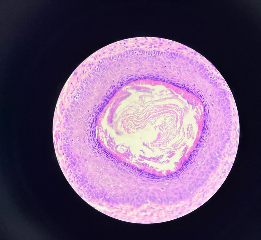
Histopathologically, numerous horn cysts are present throughout
the dermis surrounded by eosinophilic epithelial cells. In some
areas a flattened granular layer is seen between the horn cysts
and surrounding epithelial cells. We report a case of verrucous
trichoadenoma occurring on the dorsum of the nose.[1]
Case Report
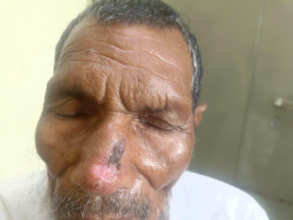
A 63‑year‑old male presented with swelling over the dorsum of
the nose for six months which began as a small blackish papule and
gradually progressed to a lesion measuring 2 × 1.5 cm.
The lesion in our patient was asymptomatic, with no associated pain, discharge, ulceration, or bleeding.
On examination, a firm non‑tender plaque measuring 2 × 1.5 cm was present over the dorsum of the nose with ill‑defined borders. Differential diagnoses considered were benign lichenoid keratosis, trichoepithelioma, trichofolliculoma, trichoadenoma, melanoma, basal cell carcinoma, pilar sheath acanthoma, and eccrine poroma.
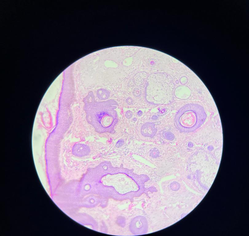
Wide local excision was performed and sent for histopathological
examination. Histopathology revealed hyperkeratosis, acanthosis and
papillomatosis of epidermis with multiple keratins filled cystically
dilated hair follicles in the dermis. Final diagnosis of verrucous
trichoadenoma was made.
Discussion
Trichoadenoma of Nikolowsky is a rare follicular tumor described
as a neoplastic process by some authors and a benign malformation
by others. It usually presents as a slow growing nodular lesion on the
face or buttocks [2]
Although trichoadenoma most frequently occurs on the face and buttocks, lesions have also been reported on the trunk, thighs and extremities, making clinical diagnosis difficult when occurring at unusual sites.
Age and Sex Predilection: Trichoadenoma typically occurs in adults in the fourth to sixth decades of life with no consistent sex predilection reported in literature. The lesion usually presents as a solitary slow‑growing papule or nodule.
The histogenesis remains uncertain but is thought to be related to trichoepithelioma and trichofolliculoma. The extensive cystification helps differentiate it from basal cell carcinoma and trichoepithelioma. Trichoadenoma is an uncommon adnexal tumor with fewer than 20 cases reported in literature, making recognition important when evaluating verrucous facial lesions.
Treatment of trichoadenoma is complete surgical excision. Recurrence: Complete surgical excision is the treatment of choice. Recurrence is uncommon when the lesion is completely removed and malignant transformation has not been reported.
This case highlights the importance of considering trichoadenoma in the differential diagnosis of verrucous lesions of the face. Histopathological examination remains essential for establishing the diagnosis and distinguishing it from malignant cutaneous tumors.
References
Citation
Bhattacharya S. Verrucous Trichoadenoma: A Case Report of a Rare Hair Follicle Tumor. Indian J Dermatol Res. 2026;4(1): 105.