


Research Article
Computed Tomography Assessment of Vascular Involvement and Diagnostic Accuracy in Pancreatic Tumors
Harshavardhan M1, Patnaik S 2*, Rammurti S1, Varma Gunturi SR3, and Uppin MS3
1Senior Resident, Department of Radiodiagnosis, Nizam’s Institute of Medical Sciences, Hyderabad, Telangana, India.
2Professor, Department of Radiodiagnosis, Nizam’s Institute of Medical Sciences, Hyderabad, Telangana, India.
3Sr Professor & Head, Department of Radiodiagnosis, Nizam’s Institute of Medical Sciences, Hyderabad, Telangana, India.
4Additional Professor. Department of Surgical Gastroenterology, Nizam’s Institute of Medical Sciences, Hyderabad, Telangana, India.
5Professor, Department of Pathology, Nizam’s Institute of Medical Sciences, Hyderabad, Telangana, India.
*Corresponding author:Dr. Sujata Patnaik, 505 A Tower, Kalpataru Residency Ashok Marg, Erragada, Hyderabad, Telangana, India. E-mail id: sujata_patnaik222@yahoo.co.in
Copyright: ©2026 Harshavardhan M, et al. This is an open access article distributed under the Creative Commons Attribution License, which permits unrestricted use, distribution, and reproduction in any medium, provided the original work is properly cited.
Article Information:Submission: 10/08/2026; Accepted: 02/10/2026; Published: 06/10/2026
Abstract
Introduction:Multi-Detector Computed Tomography (MDCT) is the most often utilised imaging modality for diagnosis, staging and resectability assessment of pancreatic tumours. Aim of this study was to evaluate the role of vascular involvement in deciding the resectability of pancreatic tumours and specify the imaging characteristics of different pancreatic tumours with special emphasis on diagnostic accuracy.
Material and Methods:All patients undergoing surgical exploration or resection for suspected pancreatic neoplasms were included in the study after ethical approval. Scans were done with 128 slice CT in pancreatic protocol. Fifty-one patients were initially screened, who were referred to the department of radiology with suspicion of pancreatic tumour.
Results:Majority of the patients were in the age group of 51-60 years. Of the 33 cases finally analysed 26 were diagnosed correctly before surgery and 7 had discordant diagnosis. 3 cases of adenocarcinoma turned out to be IPMN, NET and signet ring carcinoma on biopsy report. 1 case of NET was Castleman’s disease. Two cases of SPEN on initial assessment turned out to be Castleman’s disease, and groove pancreatitis after biopsy. One case of NET was SPEN on histopathology. Total of 264 vessels across the 33 patients were studied for accuracy of MDCT to detect vascular involvement. Of these 225 were in grade 0; 5 in grade 1; 25 in grade 2; 5 in grade 3 and 4 in grade 4 involvement with negative predictive value of 100%.
Conclusion:The value of MDCT for detection of vascular involvement in pancreatic tumours to decide on resectability was excellent with a sensitivity of 100%, specificity of 94.7 %, Positive predictive value of 77.8%, and overall diagnostic accuracy of 87.9%.
Keywords:Pancreatic cancers; Neuro Endocrine Tumours; Intraductal papillary mucinous neoplasm; Solid Pseudo papillary Epithelial Neoplasms
Introduction
Pancreatic cancer currently ranks as the 12th most common
cancer in the world and the fourth leading cause of cancer-related
deaths [1]. Early surgery is the only option for its potential cure.
However, most patients present in advanced stage with only 15- 20% of patients have resectable disease. Five- year survival after surgery is 15-27% [2]. Imaging of the pancreas is challenging because of its peculiar location in the retro peritoneum and its intricate relationship with major blood vessels and bowel. CT scan has undergone a rapid evolution over the last two decades, with each new development enhancing the imaging capability of the technique and even threatening to overwhelm our current capability to process data. Hence staging assessment for resectability is important in diagnostic imaging. Recently, the introduction of MDCT has offered a substantial improvement in volume coverage speed with no loss of image quality, facilitating optimal pancreatic parenchymal and peripancreatic vascular enhancement (6). In addition, MDCT facilitates the generation of multiplanar reconstructions, such as curved planar reformations, providing the potential to improve the detection and staging of pancreatic adenocarcinoma.
MDCT is the most commonly utilised imaging modality for diagnosis, staging and resectability assessment. Aim of this study was to assess the role of vascular involvement in deciding the resectability of pancreatic tumours and the demographic characteristics of different pancreatic tumours with special emphasis on diagnostic accuracy of pancreatic cancer.
Material and Methods
This prospective observational study was conducted over a
period of 18 months in the department of Radiodiagnosis, Nizam’s
Institute of Medical Sciences, Hyderabad. Ethical consideration: The
study protocol was subjected to clearance and approval from the
Institutional Ethics Committee (IEC). Written informed consent of
the patients or patients relatives was taken for their inclusion in the
study. Patients referred with clinical and/or sonological diagnosis or
suspected diagnoses of pancreatic neoplasms were included regardless
of the sex and age. A few unresectable cases with surgical intervention
curative surgery/palliative surgery/exploratory laparotomy done were
also included. Adenocarcinoma (known as pancreaticobiliary type)
that occurs in periampullary head region was considered as pancreatic
malignancy in our study. We excluded the patients who had surgical
or radiotherapy treatment and were already on follow up. Patients
contraindicated to the contrast study, unresectable tumours and the
metastatic pancreatic disease were excluded. The study protocol was
subjected to clearance and approval from the Institutional Ethics
Committee (IEC). A written informed consent was obtained from the
patients for their inclusion into the study. Age, sex, clinical symptoms
of the patients was recorded. Both non contrast and contrast CT
were obtained using 128 slice MDCT (SOMATOM definition AS+/
SIEMENS. Standard pancreatic protocol, NECT followed by CECT
was performed. Images obtained during pancreatic phase at 40-50sec
and portal venous phase at 65-70sec following the commencement
of IV contrast injection at an injection rate of 3.5ml/sec. Image
reconstructions and image display were done. All the images were
analysed in term of site, size, imaging morphology, local and distant
extent including vascular involvement.
Grading of vascular involvement was done according to Lu et al Grade 0 was given if there was a plane between tumor and vessel, grade 1 when the tumor–vessel contact was less than or equal to 90°, grade 2 when the tumor vessel contact was greater than 90° up to 180°, grade 3 when the tumor–vessel contact exceeded 180° but was less than 270°, and grade 4 when the tumor vessel contact was greater than or equal to 270° [3].
Grading of vascular involvement was done according to Lu et al Grade 0 was given if there was a plane between tumor and vessel, grade 1 when the tumor–vessel contact was less than or equal to 90°, grade 2 when the tumor vessel contact was greater than 90° up to 180°, grade 3 when the tumor–vessel contact exceeded 180° but was less than 270°, and grade 4 when the tumor vessel contact was greater than or equal to 270° [3].
Results
Fifty-one patients were initially screened, who were referred to
the department of radiology in suspicion of pancreatic tumour. The
age of these patients ranged from 21 to 90 years. The highest number
of patients belonged to the 51–60 years age group-13 patients (25.5%),
followed closely by the 41–50 -12 patients and 61-70 age groups-12
patients (each 23.5%). Among the total population, we found that,
twenty-eight were females (54.9%) and 23 were males (45.1%). Out
of 51 scanned cases, 18 did not undergo any surgical intervention.
However, all patients had HPE correlation. Hence the final analysis
included 33 patients.
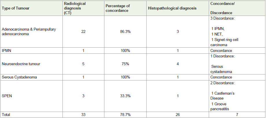
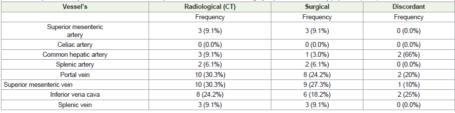
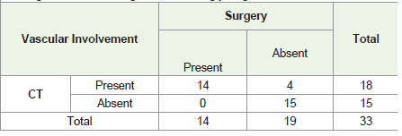
Of 33 patients of this analysis, periampullary region mass was the most common site, with 14 cases (42.4%), followed by mass in the head and uncinate process with 8 cases (24.2%). The body and head of the pancreas each were 5 cases (15.2%), while the distal body and tail region had 1 case (3.0%). Adenocarcinoma and periampullary adenocarcinoma was diagnosed in 22 patients based on CT, of which 19 (86.3 %) were confirmed histologically, resulting in an 86.3% concordance rate. The remaining 3 cases turned out to be intraductal tubulo- papillary neoplasm (ITPN), NET, and signet ring carcinoma. In the neuroendocrine tumour (NET) group, 5 cases were diagnosed radiologically, while 4 (75%) were confirmed histologically, giving a 75% concordance rate. The remaining 1 case was SCA a discordant diagnosis. For solid pseudopapillary epithelial neoplasm (SPEN), 3 cases were diagnosed radiologically, but only 1 was confirmed on histopathology, resulting in a 33.3% concordance. The two discordant cases were later identified as Castleman’s disease and groove pancreatitis, which can mimic SPEN radiologically. One case of IPMN and 1 neuroendocrine tumour were correctly diagnosed both on imaging and HPE. In total, 26 of the 33 cases (78.7%) showed concordance between CT diagnosis and histopathological findings, while 7 cases (21.3%) were discordant (Table 1). CT demonstrated strong diagnostic reliability in identifying the most common pancreatic tumours such as adenocarcinoma, with high concordance rates in several categories. Out of 33 operated cases 14 cases had both surgical and CT involvement of vessels and 4 were wrongly diagnosed on CT. Similarly, 15 were negative both on CT and surgery (Tables 2 ) (Table 3).
Total 264 vessels were studied in our 33 patients. Of these, 225 (85.2%) showed grade 0 (no involvement), 5 showed grade 1, 25 were in grade 2, five were in grade 3, four showed grade 4 (most severe) involvement. The negative predictive value (NPV) was 100% with regards to vascular involvement. The positive predictive value (PPV) was 77.8%, and the overall diagnostic accuracy of CT in detecting vascular involvement was 87.9%.
Discussion
The imaging features were critically analysed in 33 patients out
of the 51 initially screen ed. Age ranged from 21 to 90 years, and
highest number of patients were in the 51-60 years age group (25.5%).
Hossain MS et al observed males were more affected than females
and maximum were in age group of 56-65 like our study with M:
F ratio of 19:14. In their study the adeno carcinoma was the most
common variety, and the periampullary region was most common
site of involvement [4].
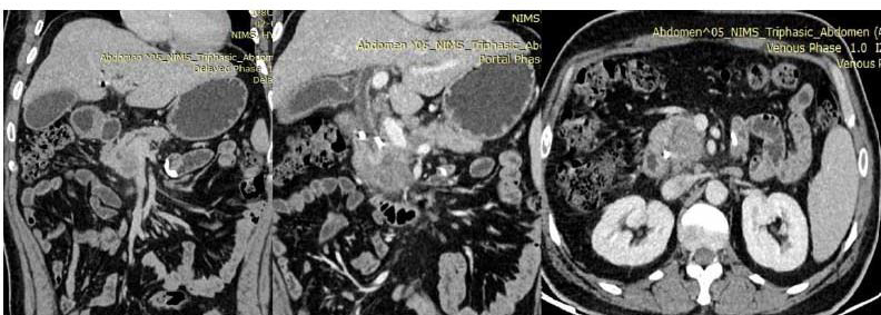
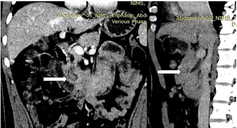
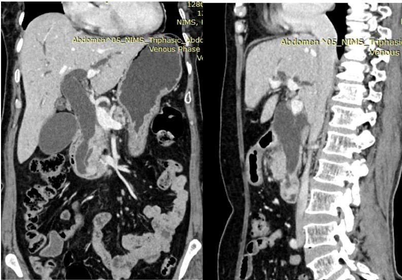
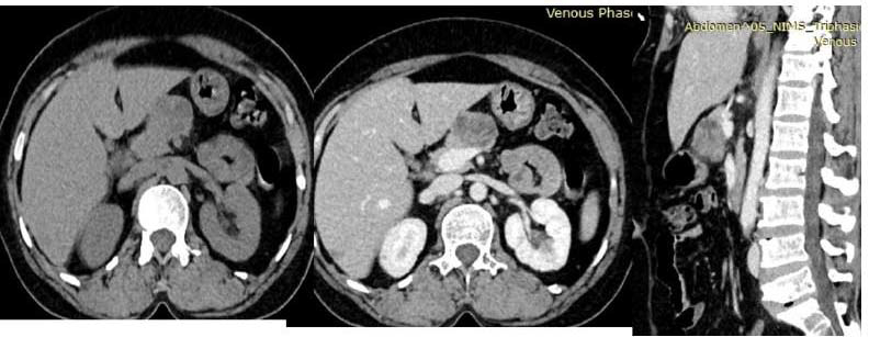
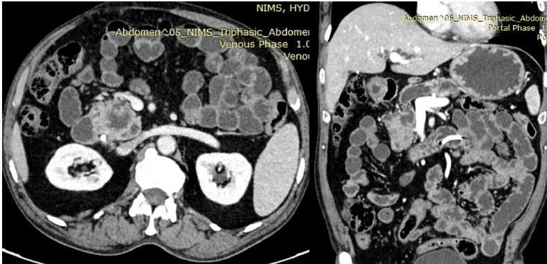
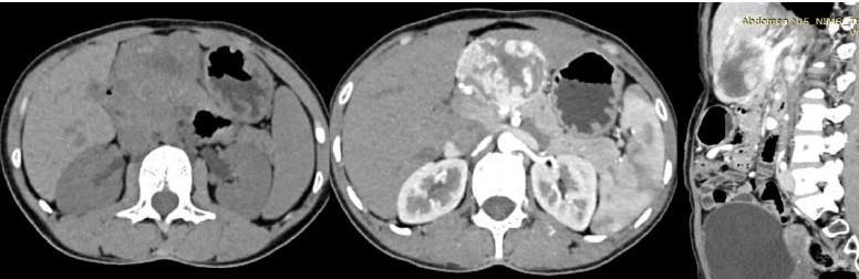
In study by Xia J et al, pancreatic adenocarcinoma accounted for 90% of all pancreatic neoplasms and most of them were above 60 years like our study (most common age group was 50-70) [Figure 1] [5]. Accurate diagnosis was possible in our study in 11 out of 14 (86%) due to distinct imaging features of hypodense mass with double ducts dilation [Figure 1]. 3 cases were thought to be adeno ca, on HPE but, they were turned out to be intra-ductal tubulepapillary neoplasm (ITPN) [Figure 2] signet ring carcinoma and NET [Figure 3]. Atypical PDAC may have intratumoral cyst due to necrosis or may accompany peritumoral non neoplastic cystic lesion or pseudocyst mimicking cystic neoplasm as seen in one of our cases [Figure 2] [6]. ITPN accounts for <1% of pancreatic neoplasm [7]. Motogusi et al [7] described double duct sign due to solid intraductal growth and seen in 70% in CT and 63% on MRI ,100% on EUS. These lesions are iso to slight hyperdense. margins are more well defined and slightly more enhancing in arterial and venous phase as compared to pancreatic adeno carcinoma [8]. Many authors reported overlapping features of ITPN, PDAC, and IPMN as our case [9]. Our case also has double duct sign positive [Figure 2]. Jeon et al [10] also reported misdiagnosis of cases of NET as adeno ca due to nonvascular NET.
In study by Xia J et al, pancreatic adenocarcinoma accounted for 90% of all pancreatic neoplasms and most of them were above 60 years like our study (most common age group was 50-70) [Figure 1] [5]. Accurate diagnosis was possible in our study in 11 out of 14 (86%) due to distinct imaging features of hypodense mass with double ducts dilation [Figure 1]. 3 cases were thought to be adeno ca, on HPE but, they were turned out to be intra-ductal tubulepapillary neoplasm (ITPN) [Figure 2] signet ring carcinoma and NET [Figure 3]. Atypical PDAC may have intratumoral cyst due to necrosis or may accompany peritumoral non neoplastic cystic lesion or pseudocyst mimicking cystic neoplasm as seen in one of our cases [Figure 2] [6]. ITPN accounts for <1% of pancreatic neoplasm [7]. Motogusi et al [7] described double duct sign due to solid intraductal growth and seen in 70% in CT and 63% on MRI ,100% on EUS. These lesions are iso to slight hyperdense. margins are more well defined and slightly more enhancing in arterial and venous phase as compared to pancreatic adeno carcinoma [8]. Many authors reported overlapping features of ITPN, PDAC, and IPMN as our case [9]. Our case also has double duct sign positive [Figure 2]. Jeon et al [10] also reported misdiagnosis of cases of NET as adeno ca due to nonvascular NET.
Signet ring ca is common in stomach. It is extremely rare in
ampulla of Vater. Sifert et al reported a case of signet ring ca out of 35
masses of ampulla of Vater carcinoma [13]. In a multi-centre study
of 170 cases of ampullary adeno ca, only one case was diagnosed as
signet ring carcinoma [14]. Blundell et al hypothesised that the origin
of ampulla of Vater signet ring ca is due to presence of gastric mucosa
or metaplasia [15]. Due to rarity of tumour, it was misdiagnosed as
adeno carcinoma in our study.
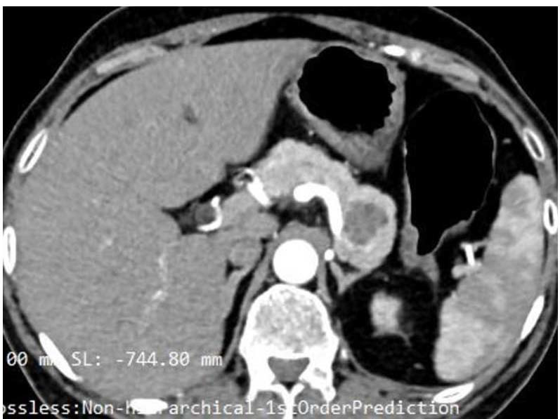
Out of 5 cases of NET, 4 were correctly diagnosed and one case was misdiagnosed as adeno ca .NET may be functional or nonfunctional.
Out of 5 cases of NET, 4 were correctly diagnosed and one case was misdiagnosed as adeno ca .NET may be functional or nonfunctional.
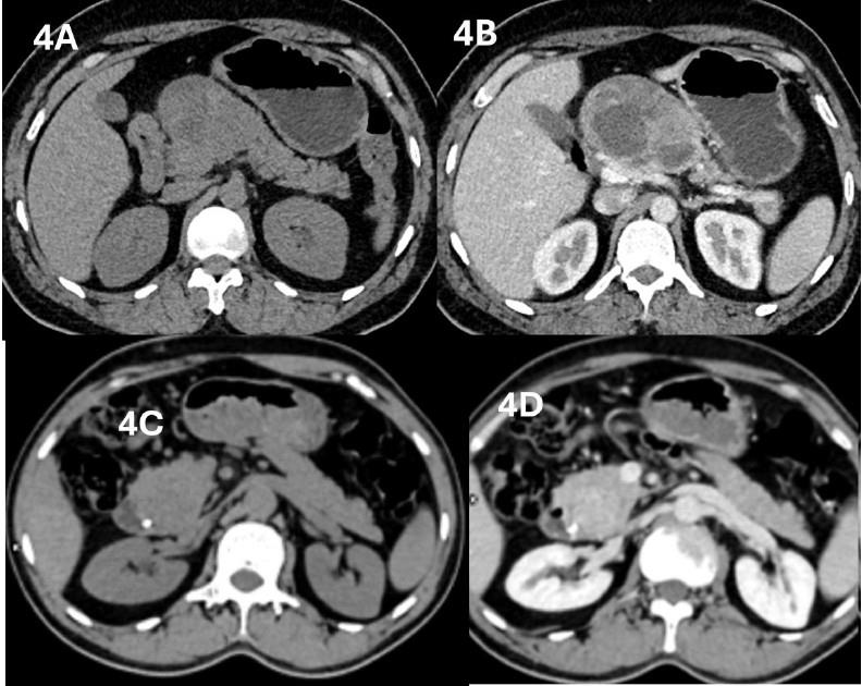
Functional tumours are less than 3cms. Non-functional tumours
are large. The typical characteristics include hypervascularity and
calcification [Figure 4]. One case was labelled as adeno ca pancreas
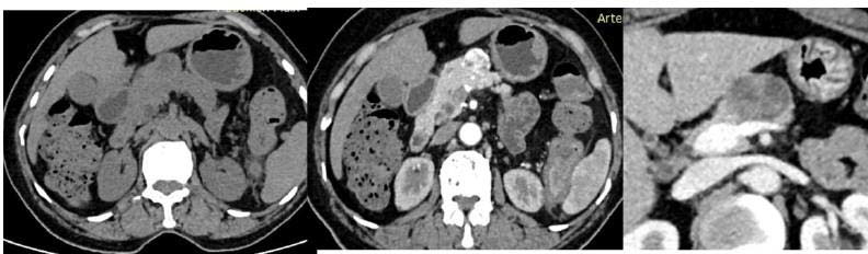
because of hypo vascularity. Sometimes NET may be atypical in
presentation in 41.5% cases and difficult to distinguish from PADC
[16]. One case was thought to be NET due to solid mass with marked
enhancement in arterial phase. HPE diagnosis was SCA [Figure 5].
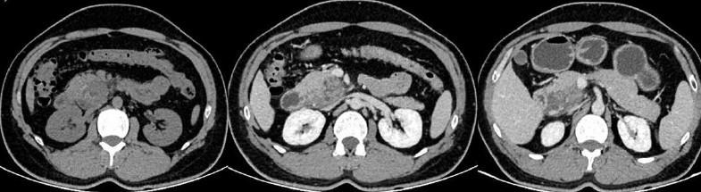
Solid pseudopapillary tumour (SPEN) is large mixed solid-cystic masses occurring in head or tail of pancreas. Tumours are well encapsulated with areas of haemorrhage and more enhancing areas in periphery and enhancement of cyst wall. We had 3 cases of SPEN; 1 was diagnosed correctly [Figure 6]. 1 case turned out to be groove pancreatitis [Figure 7] and other was histopathologically proven to be Castleman’s disease. Groove pancreatitis occurs in young adult alcoholics. Ill-defined lesion with narrowing of CBD, PD mimicking PDAC [17]. Sheet like enhancement is seen in groove pancreatitis. Cystic dystrophy of duodenal wall and smooth bile duct narrowing are other features of groove pancreatitis. Because of early age and solid cystic lesion, it was misdiagnosed as SPEN [Figure 7] in our analysis.
Solid pseudopapillary tumour (SPEN) is large mixed solid-cystic masses occurring in head or tail of pancreas. Tumours are well encapsulated with areas of haemorrhage and more enhancing areas in periphery and enhancement of cyst wall. We had 3 cases of SPEN; 1 was diagnosed correctly [Figure 6]. 1 case turned out to be groove pancreatitis [Figure 7] and other was histopathologically proven to be Castleman’s disease. Groove pancreatitis occurs in young adult alcoholics. Ill-defined lesion with narrowing of CBD, PD mimicking PDAC [17]. Sheet like enhancement is seen in groove pancreatitis. Cystic dystrophy of duodenal wall and smooth bile duct narrowing are other features of groove pancreatitis. Because of early age and solid cystic lesion, it was misdiagnosed as SPEN [Figure 7] in our analysis.
There were 2 cases of IPMN which were HPE proven. One was
diagnosed in imaging due to its typical features of cystic lesions at
head of pancreas with demonstration of communication of cystic
lesion with PD in an elderly male patient [Figure 8]. Presence of
mural nodule in cystic lesion also helps in diagnosis. One case was
misdiagnosed as adenoca [Figure 2]. A 69-year-old female had
cystic lesion in tail of pancreas with multiple small cysts less than
2 cms giving honeycomb appearance. Thin septations showed
enhancement on contrast and was typical of serous cystadenoma
(SCA) [Figure 9]. Sometimes central calcific scar may be observed in
microcystic adenoma. Density of lesion may be due to proteinaceous
fluid. Haemorrhage may be observed in some cases. WHO classified
SCA into micro and macrocystic types. Again, these two types are
further classified as 1a, 1b and 11a, 11b [18]. Type 1b is solid well
circumscribed round sponge like containing numerous tiny cyst >0.1-
1cm. They may show absence of cystic change with enhancement in
arterial phase mimicking pancreatic NET as our case [Figure 5].
Marked enhancement in arterial phase and wash out in delayed
images is described in type 1b SCA whereas in NET there is marked
enhancement in arterial phase and remains hyper attenuation in PV
phase and delayed images . Close observation of our case showed
wash out in delayed images.
A 28-year-old female had a well-defined heterogeneous solid mass in neck of pancreas showing vivid enhancement with central areas of necrosis and a possibility of SPEN was offered. HPE revealed it as Castleman’s disease [Figure 10]. Due to rarity, it was not considered even in our differential diagnosis. There was a similar case report of Castleman’s disease mimicking pancreatic NET reported by Gunda D [17].
A 28-year-old female had a well-defined heterogeneous solid mass in neck of pancreas showing vivid enhancement with central areas of necrosis and a possibility of SPEN was offered. HPE revealed it as Castleman’s disease [Figure 10]. Due to rarity, it was not considered even in our differential diagnosis. There was a similar case report of Castleman’s disease mimicking pancreatic NET reported by Gunda D [17].
In this present study we conducted comparative analysis of
vascular involvement of pancreatic tumours as assessed by CT
imaging versus intraoperative surgical findings in 33 patients. Out of
the 14 patients with vascular involvement confirmed during surgery,
CT correctly identified all 14 cases resulting in a sensitivity of 100%
[Figure 1]. CT also accurately identified 15 out of 19 cases without
vascular involvement, yielding a specificity of 78.9% [Figure 7]. There
were four false positives, where CT suggested vascular involvement
that was not confirmed during surgery [Figure 5]; but importantly,
there were no false negatives, indicating a negative predictive value
(NPV) of 100%. The positive predictive value (PPV) was 77.8%,
and the overall diagnostic accuracy of CT in detecting vascular
involvement was 87.9%.
Vargas et al conducted a study in 110 major peripancreatic vessels
in 22 resected patients with pancreatic adenocarcinoma. 108 vessels
showed no invasion on imaging and were confirmed as such during
surgery, yielding a 100% NPV and an overall accuracy of 99% for
vascular invasion that aligns with our study [18]. A similar analysis
by Phoa et al CT showed a sensitivity of 100% and a specificity of
93.3% with PPV of 75%, NPV of 100% and accuracy rate of 94.4% for
vascular involvement [19]. Lu DS et al., reported a PPV of 95%, and
a NPV of 93% with a sensitivity of 84% and specificity of 98% for the
unresectability of the vessels [3]
Despite availability of the characteristic CT features of each variety of pancreatic cancer, the CT diagnostic accuracy is only 78.7%. We identified vascular involvement in all 14 cases correctly with 100% sensitivity and a specificity of 78.9%. The positive predictive value (PPV) was 77.8%, negative predictive value (NPV) of 100% and the overall diagnostic accuracy of CT in detecting vascular involvement was 87.9%. CECT abdomen with reconstruction is a valuable noninvasive modality for the diagnosing of pancreatic tumours and assessment of the vascular involvement for resectability.
Despite availability of the characteristic CT features of each variety of pancreatic cancer, the CT diagnostic accuracy is only 78.7%. We identified vascular involvement in all 14 cases correctly with 100% sensitivity and a specificity of 78.9%. The positive predictive value (PPV) was 77.8%, negative predictive value (NPV) of 100% and the overall diagnostic accuracy of CT in detecting vascular involvement was 87.9%. CECT abdomen with reconstruction is a valuable noninvasive modality for the diagnosing of pancreatic tumours and assessment of the vascular involvement for resectability.
Conclusion
MDCT is the modality of choice for diagnosis and assessment
of resectability of pancreatic tumours. Improved volume coverage,
speed and spatial resolution along the z-axis allows 3-dimensional
reformatting images with excellent resolution. It demonstrates the
complex anatomy of pancreatic mass and the vascular involvement.
References
Citation
Harshavardhan M, Patnaik S, Rammurti S, Varma Gunturi SR, Uppin MS. Computed Tomography Assessment of Vascular Involvement and Diagnostic Accuracy in Pancreatic Tumors. Indian J Appl Radiol. 2026;12(1): 240.