


Case Report
A Silent Relic in the Pelvis: Rare Case of Ancient Pelvic Schwanomma
Siddharth Pandey, Sandeep KS, Sahana HB, Sahana A and Vidhya Rani R
Department of Radio-diagnosis, Sapthagiri Institute of Medical Sciences and Research Centre, Karnataka, Bangaluru, India
*Corresponding author: Dr. Sandeep Kumar S, Department of Radio-diagnosis, Sapthagiri Institute of Medical Sciences and Research Centre, Karnataka, Bangaluru, India. E-mail id: Sandeepkumars3006@gmail.com
Copyright: © 2026 Pandey S, et al. This is an open access article distributed under the Creative Commons Attribution License, which permits unrestricted use, distribution, and reproduction in any medium, provided the original work is properly cited.
Article Information: Submission: 27/05/2026; Accepted: 13/08/2026; Published: 15/08/2026
Abstract
Schwannomas are benign nerve sheath tumors that rarely arise in the retroperitoneum, with the ancient cystic variant being exceptionally uncommon. We report a 55-year-old male who presented with symptoms of acute calculous cholecystitis and was incidentally diagnosed with a solid cystic left iliac mass on ultrasonography of abdomen and pelvis. On further MR imaging, a well-defined, encapsulated, predominantly cystic mass was seen in the left retroperitoneum. Surgical excision was performed and histopathology confirmed the diagnosis of ancient schwannoma. Recognition of its key imaging features aids in accurate preoperative diagnosis — on ultrasound it appears as a complex cystic hypoechoic mass with posterior acoustic enhancement, and MRI reveals heterogeneous T2 hyperintense signal with a low-signal fibrous rim. Complete surgical resection remains the treatment of choice with an excellent prognosis. Awareness of this rare entity and its multimodality imaging characteristics is essential for appropriate surgical planning and avoidance of misdiagnosis.
Keywords:Ancient Schwannoma; Retroperitoneal Tumor; Cystic Schwannoma; Nerve Sheath Tumor; Pelvic Schwannoma; Ultrasonography; Cross-Sectional Imaging; Magnetic Resonance Imaging
Introduction
Schwannomas, also referred to as neuromas or neurilemmomas,
are benign, slow-growing, encapsulated tumors originating from
Schwann cells of the peripheral nerve sheath. Although they
may arise anywhere along the course of a peripheral, cranial, or
autonomic nerve, the vast majority occur in the head and neck,
extremities, and mediastinum. Retroperitoneal and pelvic locations
are distinctly uncommon, collectively accounting for less than 5% of
all schwannomas, and isolated retroperitoneal schwannomas in adult
males are exceedingly rare.
A subset of schwannomas undergoes long-standing degenerative change, producing what is termed an “ancient” schwannoma. These lesions are histologically characterised by cystic degeneration, dense hyalinisation, hemosiderin deposition, calcification, and nuclear pleomorphism—features that can mimic malignancy on both imaging and gross pathology [1,2]. Cystic change within a retroperitoneal schwannoma further broadens the differential diagnosis to include entities such as lymphangioma, cystic teratoma and pancreatic pseudocyst, among others [3,4].
A subset of schwannomas undergoes long-standing degenerative change, producing what is termed an “ancient” schwannoma. These lesions are histologically characterised by cystic degeneration, dense hyalinisation, hemosiderin deposition, calcification, and nuclear pleomorphism—features that can mimic malignancy on both imaging and gross pathology [1,2]. Cystic change within a retroperitoneal schwannoma further broadens the differential diagnosis to include entities such as lymphangioma, cystic teratoma and pancreatic pseudocyst, among others [3,4].
The radiological evaluation of retroperitoneal masses relies
principally on computed tomography (CT) and magnetic resonance
imaging (MRI). Certain imaging characteristics have been described
for retroperitoneal and pelvic schwannomas, including well-defined
encapsulation, heterogeneous signal intensity reflecting internal
degeneration, and peripheral or eggshell-like calcification on CT
[5,4]. However, none of these features are pathognomonic, and
the definitive diagnosis invariably depends on histopathological
examination following surgical excision [6,1].
Herein, we present a case of a 55-year-old male with a left-sided retroperitoneal ancient cystic schwannoma, discuss the pertinent imaging findings in the context of existing literature, and highlight the role of radiology in guiding surgical management of this rare entity.
Herein, we present a case of a 55-year-old male with a left-sided retroperitoneal ancient cystic schwannoma, discuss the pertinent imaging findings in the context of existing literature, and highlight the role of radiology in guiding surgical management of this rare entity.
Case Presentation
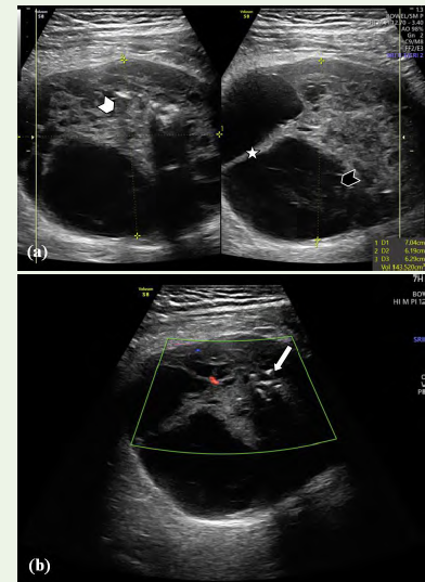
We report a case of 55-year-old male patient who presented to
us with complaints of right hypochondriac pain. On ultrasonography
of abdomen and pelvis, the imaging revealed heteroechoic collection
in the gall bladder fossa region with few hyperechoic foci within and
with discontinuity of gall bladder wall suggestive of acute calculous
cholecystitis and empyema of gall bladder. The imaging incidentally
also revealed a well-defined, solid cystic heteroechoic lesion with
internal septations and hyperechoic foci- likely calcification, in the
left iliac fossa. The lesion also shows internal vascularity on colour
doppler [Figure 1].
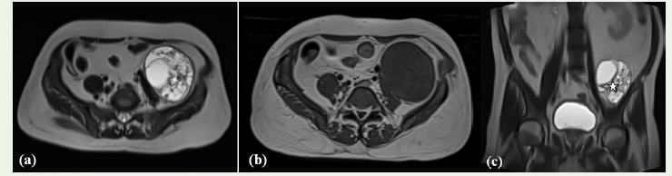
The patient was further advised for MRI abdomen and pelvis
along with MRCP, which revealed a well-defined, round to oval,
retroperitoneal encapsulated cystic lesion between the left iliacus
and psoas muscle, extending from lower border of L4 vertebra to S3
vertebral level cranio-caudally with T1 heterogenous hypointense and
T2/FLAIR hyper-intense content measuring ~ 7.6 x 7.1 x 8.9 cm (AP
x TR x CC), with T2 hypointense thick internal septations and shows
mass effect by displacing sigmoid colon anteriorly; and left psoas
major muscle, left external iliac vessels medially. There is no obvious
evidence of vascular invasion [Figure 2]
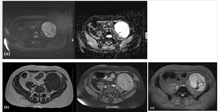
The lesion shows few areas of true diffusion restriction on
DWI and there are few T1 hyperintense foci suppressing on T1FS
suggestive of focal areas of fat and focal areas of blooming noted on
GRE imaging- s/o haemorrhage [Figure 3].
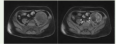
On T1 post- contrast imaging there is heterogeneous enhancement of the internal septa and solid components [Figure 4]. The lesion could not be well traced to any nerve in the present MRI.
Radiologically, the differential diagnoses of a pelvic retroperitoneal
On T1 post- contrast imaging there is heterogeneous enhancement of the internal septa and solid components [Figure 4]. The lesion could not be well traced to any nerve in the present MRI.
Radiologically, the differential diagnoses of a pelvic retroperitoneal
ancient cystic schwannoma include cystic neurogenic tumors
(neurofibroma and ganglioneuroma), tailgut cyst, lymphangioma,
mature cystic teratoma, cystic degeneration within a leiomyoma, and
necrotic malignant neoplasms such as sarcoma. Owing to extensive
degenerative changes, definitive preoperative diagnosis is often
challenging, and histopathological examination remains the gold
standard for diagnosis.
A neurogenic tumor with cystic degeneration, most likely a schwannoma, was initially suspected; however, the exact site of origin could not be determined. Exploratory laparotomy was subsequently performed through a lower midline abdominal incision. Intraoperatively, a hard solid mass was identified, causing anterior displacement of the left external iliac vessels and exerting mass effect on the left iliopsoas muscle. The lesion was meticulously dissected using a combination of blunt and sharp techniques, enabling complete excision of the pelvic mass.
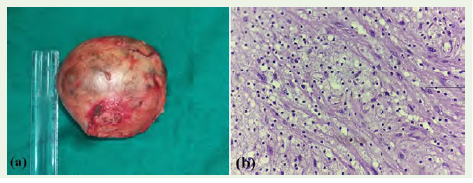
The excised mass was submitted for histopathological examination, including routine staining and sectioning. Histopathological analysis revealed a spindle-cell lesion, with features consistent with ancient schwannoma [Figure 5].
A neurogenic tumor with cystic degeneration, most likely a schwannoma, was initially suspected; however, the exact site of origin could not be determined. Exploratory laparotomy was subsequently performed through a lower midline abdominal incision. Intraoperatively, a hard solid mass was identified, causing anterior displacement of the left external iliac vessels and exerting mass effect on the left iliopsoas muscle. The lesion was meticulously dissected using a combination of blunt and sharp techniques, enabling complete excision of the pelvic mass.
The excised mass was submitted for histopathological examination, including routine staining and sectioning. Histopathological analysis revealed a spindle-cell lesion, with features consistent with ancient schwannoma [Figure 5].
Discussion
Retroperitoneal schwannomas occupy a challenging niche in
clinical radiology owing to their rarity, variable morphology, and broad
differential diagnosis. They may arise from any retroperitoneal nerve,
most commonly the lumbar and sacral nerve roots, the sympathetic
chain, or the femoral nerve, and typically follow an indolent course
with symptoms arising only when adjacent structures are compressed
[1,5]. The present case is of particular interest owing of its rarity- it
is infrequently documented in the literature—and because the tumor
demonstrated the full spectrum of ancient degenerative changes
superimposed on a cystic architecture.
The term ‘ancient’ is used to describe schwannomas that have been present in the body for a long time, resulting in secondary degenerative changes, including hyalinisation, cyst formation, calcification, hemosiderin deposition, and nuclear pleomorphism [2,3]. These changes do not indicate malignant transformation; however, the nuclear atypia seen histologically can be misinterpreted as sarcoma, underscoring the importance of radiological–pathological correlation. Macroscopic cystic change—as seen in the present case—represents one of the more dramatic manifestations of this degenerative process and is a recognised though infrequent imaging feature of ancient schwannomas [3,4].
Ultrasonography is frequently the first-line imaging modality employed in the evaluation of an abdominal or retroperitoneal mass, given its wide availability, low cost, and absence of ionizing radiation. On greyscale ultrasound, retroperitoneal schwannomas typically appear as well-defined, smoothly marginated, hypoechoic or heterogeneously echoic masses. When cystic degeneration predominates—as in the ancient variant—the lesion may demonstrate a predominantly anechoic or complex cystic echotexture with internal echoes, septations, or mural nodularity reflecting areas of residual solid Antoni tissue, hemorrhage, or hyalinisation [5,3]. Posterior acoustic enhancement is a commonly observed feature in predominantly cystic lesions and, when present, may initially suggest a simple cyst or lymphangioma [3,5]. The fibrous capsule, a hallmark of schwannomas, may be appreciated as a thin, echogenic peripheral rim on high-resolution imaging. In ancient schwannomas with significant calcification, discrete hyperechoic foci with posterior acoustic shadowing may be identified occasionally within the lesion [4]. Doppler evaluation typically reveals absent or minimal internal vascularity, with any detectable flow confined to the periphery of the solid components; this feature, combined with the well-defined margins and absence of adenopathy, helps distinguish the lesion from a vascular malformation or a hypervascular retroperitoneal neoplasm [5,1]. Despite providing valuable preliminary information, ultrasound alone is insufficient for definitive characterisation of retroperitoneal schwannomas owing to limitations in field of view, operator dependency, and restricted visualisation of deep retroperitoneal structures in adults; cross-sectional imaging with MRI is therefore essential for complete preoperative assessment [1,6].
The term ‘ancient’ is used to describe schwannomas that have been present in the body for a long time, resulting in secondary degenerative changes, including hyalinisation, cyst formation, calcification, hemosiderin deposition, and nuclear pleomorphism [2,3]. These changes do not indicate malignant transformation; however, the nuclear atypia seen histologically can be misinterpreted as sarcoma, underscoring the importance of radiological–pathological correlation. Macroscopic cystic change—as seen in the present case—represents one of the more dramatic manifestations of this degenerative process and is a recognised though infrequent imaging feature of ancient schwannomas [3,4].
Ultrasonography is frequently the first-line imaging modality employed in the evaluation of an abdominal or retroperitoneal mass, given its wide availability, low cost, and absence of ionizing radiation. On greyscale ultrasound, retroperitoneal schwannomas typically appear as well-defined, smoothly marginated, hypoechoic or heterogeneously echoic masses. When cystic degeneration predominates—as in the ancient variant—the lesion may demonstrate a predominantly anechoic or complex cystic echotexture with internal echoes, septations, or mural nodularity reflecting areas of residual solid Antoni tissue, hemorrhage, or hyalinisation [5,3]. Posterior acoustic enhancement is a commonly observed feature in predominantly cystic lesions and, when present, may initially suggest a simple cyst or lymphangioma [3,5]. The fibrous capsule, a hallmark of schwannomas, may be appreciated as a thin, echogenic peripheral rim on high-resolution imaging. In ancient schwannomas with significant calcification, discrete hyperechoic foci with posterior acoustic shadowing may be identified occasionally within the lesion [4]. Doppler evaluation typically reveals absent or minimal internal vascularity, with any detectable flow confined to the periphery of the solid components; this feature, combined with the well-defined margins and absence of adenopathy, helps distinguish the lesion from a vascular malformation or a hypervascular retroperitoneal neoplasm [5,1]. Despite providing valuable preliminary information, ultrasound alone is insufficient for definitive characterisation of retroperitoneal schwannomas owing to limitations in field of view, operator dependency, and restricted visualisation of deep retroperitoneal structures in adults; cross-sectional imaging with MRI is therefore essential for complete preoperative assessment [1,6].
MRI provides superior soft-tissue characterisation and is the
preferred modality for preoperative planning. T1-weighted sequences
demonstrate heterogeneous intermediate to low signal intensity, while
T2-weighted sequences typically reveal a markedly hyperintense
cystic component with peripheral intermediate-to-low signal from
the fibrous capsule. The so-called “target sign”—a central zone of low
T2 signal within peripheral hyperintensity—has been described in
peripheral nerve sheath tumors but is more consistently identified in
neurofibromas than schwannomas [5,6]. When present, the “split-fat
sign,” representing a rim of displaced retroperitoneal fat around the
mass, may suggest the diagnosis of a retroperitoneal schwannoma
[5]. Displacement rather than invasion of adjacent vascular and
visceral structures, and the absence of regional lymphadenopathy, are
reassuring features favouring benignity in this location [1,2].
The differential diagnosis of a left retroperitoneal cystic mass
is broad and includes lymphangioma, adrenal cyst, pancreatic
pseudocyst, cystic teratoma, Müllerian duct cyst, urachal cyst, and
cystic smooth muscle tumors [3,5]. Features that shift the differential
towards an ancient cystic schwannoma include a well-defined fibrous
capsule, a history of slow growth, absence of associated systemic
features, heterogeneous cystic architecture with solid nodularity,
peripheral calcification, and a close relationship to a named nerve on
imaging [4,6]. Biopsy, where feasible, may provide preoperative tissue
diagnosis, though surgical excision remains both therapeutic and
diagnostic [2,1].
Complete surgical resection with preservation of the parent nerve where possible is the treatment of choice and is associated with an excellent prognosis and negligible risk of malignant transformation or recurrence [1,6]. Pak and Maghsoudi emphasized the importance of identifying the nerve of origin both preoperatively on imaging and intraoperatively to minimise postoperative neurological deficits [2]. Given the retroperitoneal location and potential for sizeable tumors at presentation, multidisciplinary planning involving radiology, surgery, and oncology is advisable in complex cases [5,1].
Complete surgical resection with preservation of the parent nerve where possible is the treatment of choice and is associated with an excellent prognosis and negligible risk of malignant transformation or recurrence [1,6]. Pak and Maghsoudi emphasized the importance of identifying the nerve of origin both preoperatively on imaging and intraoperatively to minimise postoperative neurological deficits [2]. Given the retroperitoneal location and potential for sizeable tumors at presentation, multidisciplinary planning involving radiology, surgery, and oncology is advisable in complex cases [5,1].
Conclusion
Retroperitoneal ancient cystic schwannoma is an exceptionally
rare entity. This case underscores the diagnostic challenge posed by
this tumor in a non-characteristic location and patient demographic.
Radiologists should maintain familiarity with the full spectrum of
imaging appearances across modalities: on ultrasound, the lesion
typically presents as a well-defined hypoechoic or complex cystic mass
with posterior acoustic enhancement, possible peripheral echogenic
foci from calcification, and absent or minimal internal vascularity
on Doppler assessment; and on MRI, a markedly hyperintense cystic
component on T2-weighted imaging with a low-signal fibrous capsule
and moderate enhancement of solid components is characteristic.
When these features are recognised collectively, preoperative
suspicion for schwannoma can be appropriately raised. Crosssectional
imaging with MRI remains the cornerstone of definitive
lesion characterisation, operative planning, and postoperative
surveillance, with ultrasound serving as a valuable initial screening
tool. Ultimately, the combination of a thoughtful multimodality
radiological assessment and complete histopathological evaluation
enables accurate diagnosis and optimal patient outcomes in this rare
but instructive tumor type.
Patient Consent:
Written informed consent was obtained from the patient for
publication of this case report and accompanying images.References
Citation
Pandey S, Sandeep KS, Sahana HB, Sahana A,Rani RV. A Silent Relic in the Pelvis: Rare Case of Ancient Pelvic Schwanomma. Indian J Appl Radiol. 2026;12(1): 238.