


Case Report
The Mushroom Gyri Sign: MRI Diagnosis of Ulegyria as a Sequela of Perinatal Hypoxic-Ischemic Injury
Sanaulla M*, Sandeep KS, Vishwaprem RD, Krushik TR and Sugavasi GK
Department of Radiodiagnosis, Sapthagiri Institute of Medical Sciences and Research Centre, Karnataka, Bangaluru, India
*Corresponding author:Dr Mirza Sanaulla, Junior Resident, Department of Radiodiagnosis, Sapthagiri Institute of Medical Sciences and Research Centre, Karnataka, Bangaluru, India. E-mail id: mirzasanaulla14@gmail.com
Article Information:Submission: 01/06/2026; Accepted: 10/08/2026; Published: 14/08/2026
Abstract
Ulegyria is a distinctive but under-recognised form of cortical scarring that follows perinatal hypoxic-ischemic injury in the term brain. Because the depths of the sulci lie in the arterial border zones and are perfused from the pial surface inward, ischemia preferentially injures the sulcal base while sparing the gyral crown, producing atrophic convolutions with a characteristic mushroom shape. We report a 7-year-old boy, born at term with a history of failure to cry at birth and neonatal intensive care admission, who presented with global developmental delay, intellectual disability and epilepsy. Magnetic resonance imaging of the brain demonstrated brachycephaly with a simplified frontal gyral pattern, symmetrical signal hyperintensity in the perirolandic and periventricular white matter of the frontal, temporal and occipital lobes, and volume loss with atrophic, mushroom-shaped gyri in the posterior temporal, parietal and occipital watershed regions that spared the gyral crests, the hallmark of ulegyria. Secondary changes of a thinned corpus callosum and ex vacuo dilatation of the
lateral ventricles were present, while the deep grey nuclei, cerebellum and brainstem were preserved. The constellation indicated chronic sequelae of term perinatal hypoxic-ischemic injury of the partial, prolonged watershed type. Recognising ulegyria allowed the injury to be confidently ascribed to the perinatal period rather than to a later insult, and explained the child’s epilepsy and developmental impairment. Familiarity with this pattern guides counselling, supports the timing of injury, and informs management of the frequently drug-resistant epilepsy.
Keywords:Ulegyria; Mushroom Gyri; Hypoxic-Ischemic Encephalopathy; Watershed Injury; Epilepsy.
Introduction
Ulegyria is a form of cortical scarring that follows hypoxicischemic
injury in the term or near-term brain; the name derives
from the Greek ule, meaning scar.[1] Its defining feature is atrophy
concentrated at the depths of the sulci with relative preservation of
the gyral crowns, so that the affected convolutions take on a narrow stemmed,
mushroom shape.[1,2] This topography reflects cortical
vascular anatomy: the sulcal depths lie within the arterial border zones
and are perfused from the pial surface inward, making the sulcal base a
watershed within the watershed that is selectively vulnerable to falling
perfusion.[2,3] Ulegyria is therefore characteristic of the partial,
prolonged pattern of term injury, which targets the parasagittal
cortex and subcortical white matter, in contrast to the periventricular
leukomalacia of the preterm infant.[2,3] It is strongly associated with
cerebral palsy, intellectual disability and, especially, drug-resistant
epilepsy, yet is frequently overlooked on delayed imaging.
[1,4,5] We report a child in whom magnetic resonance imaging (MRI) showed
classical ulegyria as the sequela of term perinatal asphyxia. Beyond
illustrating the classical appearance, we use the case to make explicit a
connection that is easy to overlook at the reporting console: ulegyria
and the Dyke-Davidoff-Masson syndrome are not separate diagnoses
but two outcomes of a single early insult, set apart chiefly by whether
the damage was symmetrical or lateralised, and we draw attention
to the calvarial remodelling that accompanies an early, global loss of
cerebral volume.
Case Report
A 7-year-old boy presented with global developmental delay
affecting speech, language and motor milestones, poor social skills,
intellectual disability and epilepsy. He had been born at term but
did not cry at birth and required neonatal intensive care admission;
earlier records were unavailable. MRI of the brain was performed.
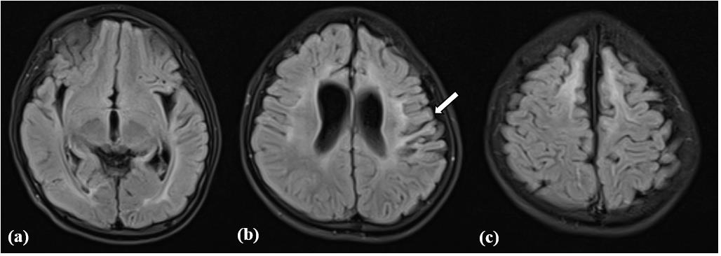
Axial FLAIR images showed symmetrical subcortical and periventricular white-matter hyperintensity in the temporo-occipital, parietal and frontal lobes, with marked volume loss of the adjacent white matter. In the parietal watershed region, the overlying gyri were small and atrophic and spared the gyral crests, giving the characteristic mushroom-shaped configuration of ulegyria, with ex vacuo dilatation of the lateral ventricles [Figure 1].
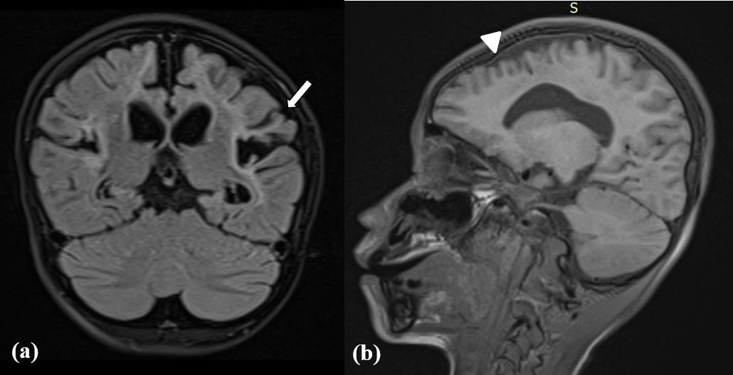
On the coronal plane the same mushroom-shaped gyri sparing the gyral crests were confirmed, with increased FLAIR signal and volume loss in the subcortical white matter of the parietal lobes, while the sagittal T1-weighted image showed a simplified frontal gyral pattern [Figure 2].
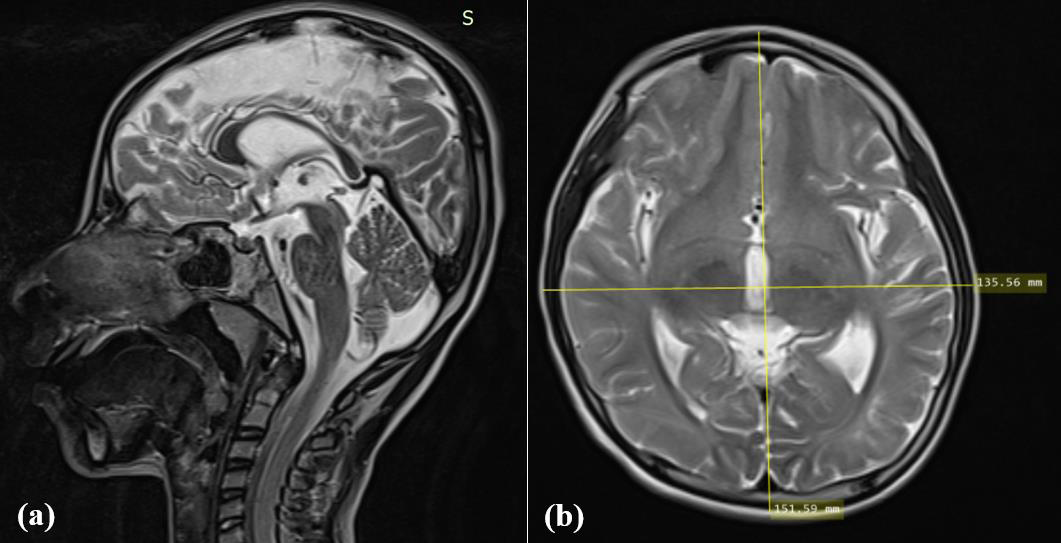
The midline sagittal T2-weighted image showed thinning of the body and splenium of the corpus callosum, and the axial T2-weighted image gave a cephalic index of 89 (135.56 by 151.59 mm), confirming brachycephaly [Figure 3]. The basal ganglia, thalami, cerebellum, brainstem and posterior fossa were normal, with no acute infarct, hemorrhage or mass. These appearances were diagnostic of chronic sequelae of term perinatal hypoxic-ischemic injury of the partial, prolonged watershed type, with ulegyria.
Axial FLAIR images showed symmetrical subcortical and periventricular white-matter hyperintensity in the temporo-occipital, parietal and frontal lobes, with marked volume loss of the adjacent white matter. In the parietal watershed region, the overlying gyri were small and atrophic and spared the gyral crests, giving the characteristic mushroom-shaped configuration of ulegyria, with ex vacuo dilatation of the lateral ventricles [Figure 1].
On the coronal plane the same mushroom-shaped gyri sparing the gyral crests were confirmed, with increased FLAIR signal and volume loss in the subcortical white matter of the parietal lobes, while the sagittal T1-weighted image showed a simplified frontal gyral pattern [Figure 2].
The midline sagittal T2-weighted image showed thinning of the body and splenium of the corpus callosum, and the axial T2-weighted image gave a cephalic index of 89 (135.56 by 151.59 mm), confirming brachycephaly [Figure 3]. The basal ganglia, thalami, cerebellum, brainstem and posterior fossa were normal, with no acute infarct, hemorrhage or mass. These appearances were diagnostic of chronic sequelae of term perinatal hypoxic-ischemic injury of the partial, prolonged watershed type, with ulegyria.
Discussion
The pattern of perinatal hypoxic-ischemic injury depends on the
maturity of the brain and on the severity and duration of the insult.
In the term infant a partial but prolonged reduction in perfusion
preferentially injures the parasagittal cortex and subcortical white
matter of the arterial border zones, whereas a profound, abrupt
insult targets the deep grey nuclei and perirolandic cortex.[2,3] The
sulcal-depth scarring that defines ulegyria follows directly from the
architecture of the cortical blood supply: because the pial branches
run inward from the surface, the floor of each sulcus is perfused last
and therefore fails first when pressure falls; as the injury matures
these undernourished stems shrink while the better-supplied
crowns retain their bulk, so that each convolution is left with the
narrow-waisted, mushroom silhouette.[2,3] In the present case, the
watershed distribution, the preserved basal ganglia and thalami, and
the perirolandic signal together indicate a predominantly partial,
prolonged injury.
The secondary features here are equally typical. Loss of cortical
and subcortical neurons leads to deafferentation and Wallerian
degeneration, manifesting as a thinned corpus callosum, while
parenchymal volume loss produces ex vacuo dilatation of the lateral
ventricles.[2] Ulegyria is best appreciated on T2-weighted and
FLAIR images as small, atrophic gyri with broad intervening sulci
and underlying white-matter gliosis, most often in the posterior and
perisylvian watershed zones; it is reported in roughly two-thirds of
children with term hypoxic-ischemic injury and cortical damage,
yet is frequently missed.[2,4] Its recognition lets the radiologist
ascribe the injury specifically to the perinatal period rather than to
a later event such as prolonged seizure, cardiac arrest or drowning, a
distinction with clinical and medicolegal weight.[4]
Clinically, ulegyria is closely linked to drug-resistant epilepsy,
cerebral palsy and cognitive impairment, with seizures often arising
from the posterior cortex and a frequent history of perinatal asphyxia,
a profile matched by our patient. [1,5,6] The chief practical value of
the mushroom-gyri sign is that it separates ulegyria from its mimics.
Foremost among these is polymicrogyria, a malformation of cortical
development in which the cortex is irregular and infolded from
the outset; unlike ulegyria it is not selectively scarred at the sulcal
depths, does not spare the gyral crowns in the way that generates the
mushroom outline, and lacks the watershed white-matter gliosis that
betrays an ischemic origin.[2,7] Focal cortical dysplasia and other
malformations of cortical development are likewise congenital and
do not follow the parasagittal, arterial border-zone distribution or
carry the secondary stigmata of remote injury — a thinned corpus
callosum, Wallerian degeneration and ex vacuo ventriculomegaly
— that attend ulegyria. Because these mimics are developmental
rather than acquired, reading the mushroom morphology together
with its watershed setting is precisely what lets the radiologist label
the process a scar rather than a malformation and, in turn, anchor it
to the perinatal period. Management is multidisciplinary; refractory
seizures may respond to antiseizure medication or, in selected cases,
to resective epilepsy surgery.[5,6]
A closely related sequela of early-life brain injury is the Dyke- Davidoff-Masson syndrome (DDMS), or cerebral hemiatrophy, which shares with ulegyria an origin in an ischemic, infective or hemorrhagic insult to the immature brain, frequently of perinatal hypoxic-ischemic type.[8] In DDMS the injury is predominantly unilateral, producing atrophy of one cerebral hemisphere with ex vacuo dilatation of the ipsilateral lateral ventricle and, when the insult occurs before about three years of age, compensatory osseous changes — ipsilateral calvarial (diploic) thickening, hyperpneumatization of the frontal, ethmoid and sphenoid sinuses and the mastoid air cells, and elevation of the petrous ridge and sphenoid wing — as the atrophic hemisphere exerts diminished outward pressure on the still-developing calvarium.[8] The same principle operated in our patient: the global reduction in cerebral volume was accompanied by a calvarial response, but because the injury was bilateral and symmetrical it produced brachycephaly rather than the unilateral hemicranial hypertrophy of DDMS, and the cortical scarring took the watershed, mushroom-gyri form of ulegyria rather than frank hemiatrophy. Recognising this distinction keeps the lateralising hemiatrophy and asymmetric skull changes of DDMS separate from the symmetrical parasagittal scarring of ulegyria, even though both lie on the spectrum of remote perinatal hypoxic-ischemic injury.
A closely related sequela of early-life brain injury is the Dyke- Davidoff-Masson syndrome (DDMS), or cerebral hemiatrophy, which shares with ulegyria an origin in an ischemic, infective or hemorrhagic insult to the immature brain, frequently of perinatal hypoxic-ischemic type.[8] In DDMS the injury is predominantly unilateral, producing atrophy of one cerebral hemisphere with ex vacuo dilatation of the ipsilateral lateral ventricle and, when the insult occurs before about three years of age, compensatory osseous changes — ipsilateral calvarial (diploic) thickening, hyperpneumatization of the frontal, ethmoid and sphenoid sinuses and the mastoid air cells, and elevation of the petrous ridge and sphenoid wing — as the atrophic hemisphere exerts diminished outward pressure on the still-developing calvarium.[8] The same principle operated in our patient: the global reduction in cerebral volume was accompanied by a calvarial response, but because the injury was bilateral and symmetrical it produced brachycephaly rather than the unilateral hemicranial hypertrophy of DDMS, and the cortical scarring took the watershed, mushroom-gyri form of ulegyria rather than frank hemiatrophy. Recognising this distinction keeps the lateralising hemiatrophy and asymmetric skull changes of DDMS separate from the symmetrical parasagittal scarring of ulegyria, even though both lie on the spectrum of remote perinatal hypoxic-ischemic injury.
Framed in this way, the present case offers more than another
example of an uncommon sign. It shows the two ends of a single
continuum meeting in one patient — the parasagittal, mushroomgyri
scarring of ulegyria alongside the whole-skull remodelling
that, when an insult happens to be unilateral, declares itself as the
Dyke-Davidoff-Masson syndrome. For the reporting radiologist the
practical yield is threefold: the mushroom-gyri sign fixes the timing
of the injury within the perinatal window, the symmetry of both the
cortical and the calvarial changes distinguishes a global insult from
a lateralised one, and the recognition itself matters because ulegyria
continues to be under-reported and its posterior-cortex epilepsy,
though often drug-resistant, is frequently amenable to resective
surgery.[9]
Conclusion
Ulegyria is the imaging signature of remote term perinatal
hypoxic-ischemic injury. The combination of atrophic, mushroom shaped
gyri sparing the gyral crests in the parasagittal watershed
zones, white-matter gliosis, a thinned corpus callosum and ex vacuo
ventriculomegaly, with preserved deep grey nuclei, allows the injury
to be confidently dated to the perinatal period and explains the
associated epilepsy and developmental impairment. Familiarity with
this pattern supports accurate diagnosis, appropriate counselling and
timely management of the often-drug-resistant epilepsy.
Learning Points
1. Ulegyria is the chronic imaging signature of term, partialprolonged
perinatal hypoxic-ischemic injury; its hallmark is atrophy
at the sulcal depths with sparing of the gyral crowns, producing
mushroom-shaped gyri in the parasagittal watershed zones that are
best appreciated on T2-weighted and FLAIR images.
2. Identifying the sign allows the injury to be dated to the perinatal period rather than attributed to a later event such as prolonged seizure, cardiac arrest or drowning, a distinction that carries both clinical and medicolegal weight.
3. Ulegyria and the Dyke-Davidoff-Masson syndrome lie on one spectrum of remote perinatal injury; symmetry and laterality determine whether the result is bilateral mushroom-gyri with brachycephaly or unilateral hemiatrophy with hemicranial osseous
2. Identifying the sign allows the injury to be dated to the perinatal period rather than attributed to a later event such as prolonged seizure, cardiac arrest or drowning, a distinction that carries both clinical and medicolegal weight.
3. Ulegyria and the Dyke-Davidoff-Masson syndrome lie on one spectrum of remote perinatal injury; symmetry and laterality determine whether the result is bilateral mushroom-gyri with brachycephaly or unilateral hemiatrophy with hemicranial osseous
overgrowth.
4. Because ulegyria-related epilepsy is frequently posterior in origin and drug-resistant yet may respond to resective surgery, flagging the pattern has direct value for management and counselling.
Patient Consent
Written informed consent for publication of this case report and the accompanying images was obtained from the patient’s parent/ guardian.
4. Because ulegyria-related epilepsy is frequently posterior in origin and drug-resistant yet may respond to resective surgery, flagging the pattern has direct value for management and counselling.
Patient Consent
Written informed consent for publication of this case report and the accompanying images was obtained from the patient’s parent/ guardian.
Conflict of Interest:
The authors declare no conflict of interest.References
Citation
Sanaulla M, Sandeep KS, Vishwaprem RD, Krushik TR, Sugavasi GK. The Mushroom Gyri Sign: MRI Diagnosis of Ulegyria as a Sequela of Perinatal Hypoxic-Ischemic Injury. Indian J Appl Radiol. 2026;12(1): 237.